The cell membrane serves as a dynamic barrier that regulates what enters and exits the cell, composed primarily of a phospholipid bilayer with embedded proteins. This diagram highlights the structure of the membrane and the critical role of transmembrane proteins, including ion channel proteins that facilitate the movement of ions across the membrane. Understanding these components offers valuable insights into cellular function and communication, forming the foundation of many physiological processes.
Key Labels in the Cell Membrane Diagram
This section provides an in-depth look at each labeled element, revealing their roles in cellular structure and function.
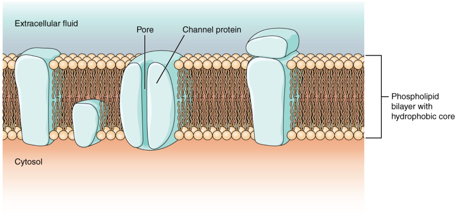
Phospholipid bilayer: This double layer of phospholipids forms the basic structure of the cell membrane, with hydrophilic heads facing outward and hydrophobic tails inward. It provides a selective barrier that controls the passage of substances into and out of the cell.
Hydrophilic head: The polar, water-attracting portion of phospholipids, located on the outer and inner surfaces of the bilayer. It interacts with the aqueous environments inside and outside the cell, stabilizing the membrane structure.
Hydrophobic tail: The nonpolar, water-repelling portion of phospholipids, situated in the interior of the bilayer. This region prevents the free diffusion of water-soluble molecules, enhancing the membrane’s selective permeability.
Transmembrane protein: These proteins span the entire bilayer, serving as channels, receptors, or transporters, such as ion channels. They facilitate communication and transport across the membrane, playing a key role in cellular signaling.
Ion channel protein: A subtype of transmembrane protein that forms pores allowing specific ions, like sodium or potassium, to pass through the membrane. Its selective nature regulates electrical impulses and maintains cellular homeostasis.
Extracellular space: The area outside the cell where the hydrophilic heads of the bilayer are exposed to the external environment. It is where signaling molecules and nutrients interact with the cell membrane.
Cytoplasm: The internal fluid environment of the cell, containing the inner layer of hydrophilic heads and various organelles. It serves as the site where transported ions and molecules exert their effects.
Peripheral protein: These proteins are attached to the inner or outer surface of the bilayer, often involved in structural support or signaling. They do not span the membrane but anchor other components.
Structure of the Cell Membrane
The cell membrane is a fluid mosaic of lipids and proteins. Its structure supports a variety of cellular functions.
- The phospholipid bilayer forms a flexible yet stable barrier.
- Hydrophilic heads interact with water, while hydrophobic tails create a hydrophobic core.
- Transmembrane proteins embed within this bilayer, enabling transport and signaling.
- The fluid nature allows lateral movement of components.
- Cholesterol within the bilayer regulates fluidity and stability.
Functions of Transmembrane Proteins
Transmembrane proteins are essential for cellular activities. They mediate critical interactions and transport.
- Ion channel proteins allow selective ion passage, crucial for nerve impulses.
- These proteins act as receptors, binding hormones or neurotransmitters.
- They facilitate the transport of glucose and amino acids.
- Some serve as anchors, linking the membrane to the cytoskeleton.
- Mutations can impair function, affecting diseases like cystic fibrosis.
Role of the Phospholipid Bilayer
The phospholipid bilayer is the foundation of membrane integrity. It balances permeability and protection.
- Hydrophilic heads ensure compatibility with aqueous environments.
- Hydrophobic tails prevent unwanted molecule passage.
- The bilayer self-assembles, maintaining cellular compartmentalization.
- It adapts to temperature changes via lipid composition.
- Damage to this structure can compromise cell viability.
Cellular Compartments and Their Interactions
The extracellular space and cytoplasm define the membrane’s boundaries. They influence cellular processes.
- The extracellular space hosts signaling molecules that bind transmembrane proteins.
- Cytoplasm contains enzymes and ions regulated by ion channel proteins.
- These compartments maintain electrochemical gradients.
- Transport across the membrane affects cytoplasmic composition.
- Imbalances can lead to osmotic stress or cell swelling.
Physiological Significance and Clinical Insights
The cell membrane’s anatomy has broad physiological implications. It influences health and disease management.
- Ion channel protein dysfunction is linked to epilepsy and cardiac arrhythmias.
- The phospholipid bilayer’s fluidity affects drug delivery and membrane repair.
- Transmembrane proteins are targets for therapies like monoclonal antibodies.
- Peripheral protein abnormalities can disrupt cell adhesion.
- Research into membrane dynamics aids in developing treatments.
In conclusion, the cell membrane and transmembrane proteins diagram illustrates a sophisticated cellular structure, with the phospholipid bilayer providing a selective barrier and transmembrane proteins enabling vital functions. From ion channel proteins regulating ion flow to the interplay between extracellular space and cytoplasm, this system underpins cellular health. Exploring these elements enhances our understanding of cellular physiology and its clinical relevance.

