Lymph nodes are vital components of the immune system, acting as filters for lymph fluid and sites for immune cell activation. These small, bean-shaped structures are strategically located along lymphatic vessels, playing a key role in detecting and responding to pathogens such as bacteria and viruses. Their intricate histology, as depicted in this micrograph, reveals a complex network of cells and tissues that collaborate to maintain lymphatic health and immunity.
Labeled Components of the Lymph Node
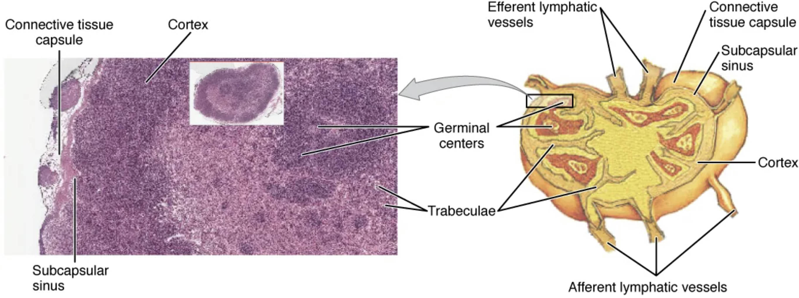
Connective tissue capsule: This outer layer encases the lymph node, providing structural integrity and protection against external damage. It is composed of dense collagen fibers that also anchor the node to surrounding tissues.
Cortex: The cortex forms the outer region of the lymph node, rich in lymphocytes and containing primary and secondary follicles. It is the primary site for initiating immune responses, particularly involving B-cell activation.
Subcapsular sinus: Located just beneath the connective tissue capsule, this sinus collects lymph fluid entering the node and directs it toward the cortex. It serves as an initial filtration point, allowing macrophages to engulf pathogens or debris.
Germinal centers: These are specialized areas within the cortex where B cells rapidly proliferate and differentiate into plasma cells or memory B cells. They are critical for humoral immunity, producing antibodies to combat specific antigens.
Trabeculae: These inward extensions of the connective tissue capsule divide the lymph node into compartments, providing support and channeling lymph flow. They also facilitate the distribution of immune cells and nutrients.
Afferent lymphatic vessels: These vessels carry lymph from peripheral tissues into the lymph node, delivering antigens and immune cells for processing. They enter the node at multiple points, ensuring thorough filtration.
Efferent lymphatic vessels: These vessels exit the lymph node, transporting filtered lymph and activated immune cells toward the bloodstream or other nodes. Typically, there is only one efferent vessel per node, located at the hilum.
Connective tissue capsule: This structure also supports the internal architecture, extending trabeculae to maintain the node’s shape and function. It plays a role in anchoring blood vessels that supply the node.
Anatomical Overview of the Lymph Node
The lymph node’s structure is a testament to its role in immune surveillance, with distinct regions working in harmony.
- The connective tissue capsule forms a protective barrier, with trabeculae extending inward to create a scaffold.
- The cortex houses lymphoid follicles, where germinal centers form in response to antigenic stimulation.
- The subcapsular sinus acts as a gateway, filtering lymph as it enters and distributing it to cortical regions.
- Afferent lymphatic vessels bring unfiltered lymph, rich in antigens, into the node for processing.
- Efferent lymphatic vessels ensure the exit of lymph after immune cells have been activated.
- Blood vessels within the node, supported by trabeculae, supply oxygen and nutrients to sustain immune activity.
This histological view, magnified at 128x, highlights the germinal centers, showcasing active B-cell division.
Physiological Functions of the Lymph Node
The lymph node is a dynamic hub for immune cell interaction, orchestrating both innate and adaptive responses.
- Lymph enters via afferent vessels, where macrophages in the subcapsular sinus begin phagocytosis of pathogens.
- In the cortex, B cells within germinal centers undergo somatic hypermutation to enhance antibody affinity.
- T cells in the paracortical areas interact with dendritic cells, initiating cell-mediated immunity.
- Efferent vessels carry activated lymphocytes and antibodies to systemic circulation.
- The connective tissue capsule and trabeculae provide stability, allowing efficient lymph flow and cell migration.
- Hormonal signals, though not primary, can modulate immune activity within the node.
This process ensures rapid response to infections, with germinal centers amplifying antibody production.
Histological Features and Cellular Dynamics
The micrograph reveals the lymph node’s cellular diversity, essential for its immunological role.
- The cortex is densely packed with lymphocytes, with germinal centers showing mitotic figures of B cells.
- Subcapsular sinuses contain macrophages and dendritic cells, key players in antigen presentation.
- Trabeculae support reticular fibers that form a network for lymphocyte movement.
- Afferent vessels deliver lymph, triggering immune activation in the cortex and medulla.
- Efferent vessels export mature immune cells, completing the lymph node’s filtration cycle.
- The connective tissue capsule maintains structural integrity, protecting against mechanical stress.
This detailed histology underscores the node’s efficiency in immune education and response.
Clinical Relevance of Lymph Node Structure
Understanding lymph node anatomy aids in diagnosing and managing immune-related conditions.
- Enlarged nodes may indicate infection or, in rare cases, lymphoma, with germinal center hyperactivity as a clue.
- The cortex’s role in B-cell maturation makes it a target for studying autoimmune diseases.
- Afferent vessel blockages can lead to lymphedema, highlighting their importance in lymph drainage.
- Efferent vessel function ensures systemic immune support, critical post-vaccination.
- Trabeculae and the capsule provide a framework, with changes signaling chronic inflammation.
- Histological analysis of germinal centers aids in assessing immune competence.
This knowledge is invaluable for interpreting biopsy results and planning treatments.
The lymph node’s intricate structure, as illustrated in this micrograph, reflects its critical role in immunity. By filtering lymph and activating immune cells, it ensures the body remains protected against a wide array of pathogens, making it a cornerstone of health that deserves closer study and appreciation.

