The lymphatic capillaries form the initial network of the lymphatic system, playing a critical role in collecting excess fluid and waste from tissues. This detailed diagram provides an insightful look into their structure and how they interact with surrounding tissues to maintain fluid balance and support immune function.
Key Labeled Components in the Lymphatic Capillaries Diagram
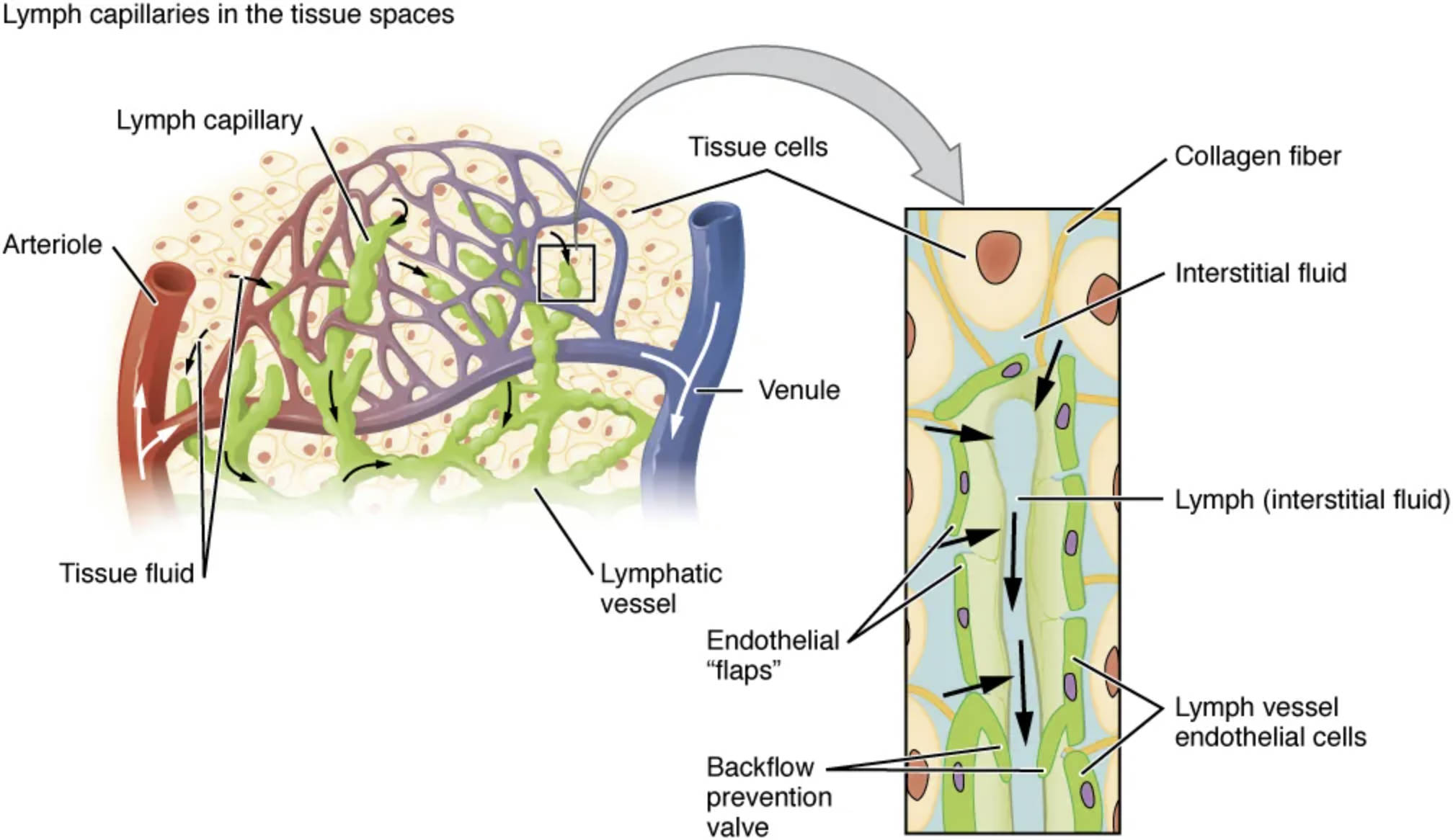
Lymph capillary The lymph capillary is a tiny, thin-walled vessel that begins the lymphatic drainage process by absorbing interstitial fluid from tissue spaces. Its unique structure with overlapping endothelial cells allows easy entry of fluid, proteins, and pathogens, initiating the journey toward larger lymphatic vessels.
Arteriole The arteriole is a small artery that delivers oxygenated blood to the tissue, contributing to the fluid that becomes interstitial fluid. Its proximity to lymph capillaries facilitates the exchange of substances, supporting tissue health and lymphatic function.
Venule The venule is a small vein that collects deoxygenated blood from the tissue, working alongside arterioles to maintain circulation. It interacts with the lymphatic system by allowing excess fluid to be drained into lymph capillaries, preventing tissue swelling.
Lymphatic vessel The lymphatic vessel transports lymph away from the capillary network toward lymph nodes for filtration. It features valves, as shown in the inset, to ensure unidirectional flow and prevent backflow of fluid.
Tissue cells Tissue cells are the living components of the tissue, surrounded by interstitial fluid that nourishes them. These cells release waste into the fluid, which is then collected by lymph capillaries to maintain a healthy environment.
Tissue fluid Tissue fluid, or interstitial fluid, is the extracellular fluid that bathes tissue cells, derived from blood plasma leaking through capillaries. It becomes lymph when absorbed by lymphatic capillaries, carrying nutrients, waste, and immune cells.
Endothelial “flaps” Endothelial “flaps” are the overlapping edges of endothelial cells that form the walls of lymph capillaries, allowing fluid to enter easily. These flaps act as one-way gates, opening to admit interstitial fluid while preventing its escape back into tissues.
Backflow prevention valve The backflow prevention valve, visible in the lymphatic vessel inset, ensures that lymph flows only toward the heart, avoiding pooling in tissues. This mechanism is crucial for maintaining efficient lymphatic circulation.
Collagen fiber The collagen fiber anchors the lymphatic capillary within the tissue, providing structural support. It also helps regulate the capillary’s openness, allowing fluid entry while maintaining stability.
Interstitial fluid Interstitial fluid fills the spaces between tissue cells, serving as a medium for nutrient delivery and waste removal. It is continuously collected by lymph capillaries to prevent accumulation and edema.
Lymph (interstitial fluid) Lymph (interstitial fluid) is the fluid once it enters the lymphatic system, carrying proteins, lipids, and immune cells. This fluid is transported through lymphatic vessels to be filtered and returned to the bloodstream.
Lymph vessel endothelial cells Lymph vessel endothelial cells line the inner walls of lymphatic vessels, facilitating smooth lymph flow. These cells also play a role in immune cell migration, supporting the body’s defense mechanisms.
Structure and Function of Lymphatic Capillaries
Lymphatic capillaries are the starting point of the lymphatic drainage system. Their unique design ensures efficient fluid uptake from tissues.
- Capillary Network: The intricate web of lymph capillaries, interlaced with arterioles and venules, maximizes fluid collection across various tissue types.
- Fluid Absorption: These capillaries absorb approximately 3 liters of interstitial fluid daily, preventing edema and maintaining tissue pressure.
- Immune Role: They collect antigens and pathogens, transporting them to lymph nodes for immune processing.
The presence of endothelial “flaps” allows selective entry of larger molecules, unlike blood capillaries. This adaptability supports the lymphatic system’s role in both fluid balance and immunity.
Microscopic Anatomy and Anchoring Mechanism
The microscopic view reveals the capillary’s delicate structure. Collagen fibers provide essential support for this network.
- Endothelial Overlap: The overlapping endothelial cells create a semi-permeable barrier, opening under pressure to admit fluid and closing to retain it.
- Collagen Support: Anchored by collagen fibers, the capillaries remain stable yet flexible, adapting to tissue movement and fluid dynamics.
- Valve Function: The backflow prevention valve in larger vessels ensures lymph moves toward lymph nodes, enhancing circulation efficiency.
This anchoring mechanism prevents collapse under tissue pressure, ensuring consistent function. The interplay between structure and fluid dynamics is key to lymphatic health.
Integration with the Cardiovascular System
Lymphatic capillaries work closely with the cardiovascular system. This collaboration is vital for overall homeostasis.
Arterioles and venules release fluid that becomes interstitial fluid, which lymph capillaries then collect. The process supports blood volume regulation by returning fluid via the thoracic duct to the venous system near the heart.
- Fluid Exchange: Approximately 90% of filtered plasma returns to venules, with the remaining 10% entering lymph capillaries, balancing circulation.
- Nutrient Transport: Lipids absorbed in the gut enter lymph as chylomicrons, bypassing initial blood filtration for efficient metabolism.
- Immune Coordination: Antigens carried in lymph trigger responses in lymph nodes, linking lymphatic and immune systems.
This integration highlights the lymphatic system’s role in supporting cardiovascular and immune functions.
In conclusion, lymphatic capillaries are fundamental to the lymphatic system, efficiently managing fluid balance and immune defense. Their anatomical design, supported by collagen and valves, ensures seamless operation with the cardiovascular system, promoting overall health and resilience.

