Venules are the smallest veins in the circulatory system, serving as the initial collectors of blood from capillaries and transitioning it toward larger veins. This image provides a detailed sectional view of a venule, revealing its microscopic structure and the layers that support its role in facilitating blood flow and exchange.
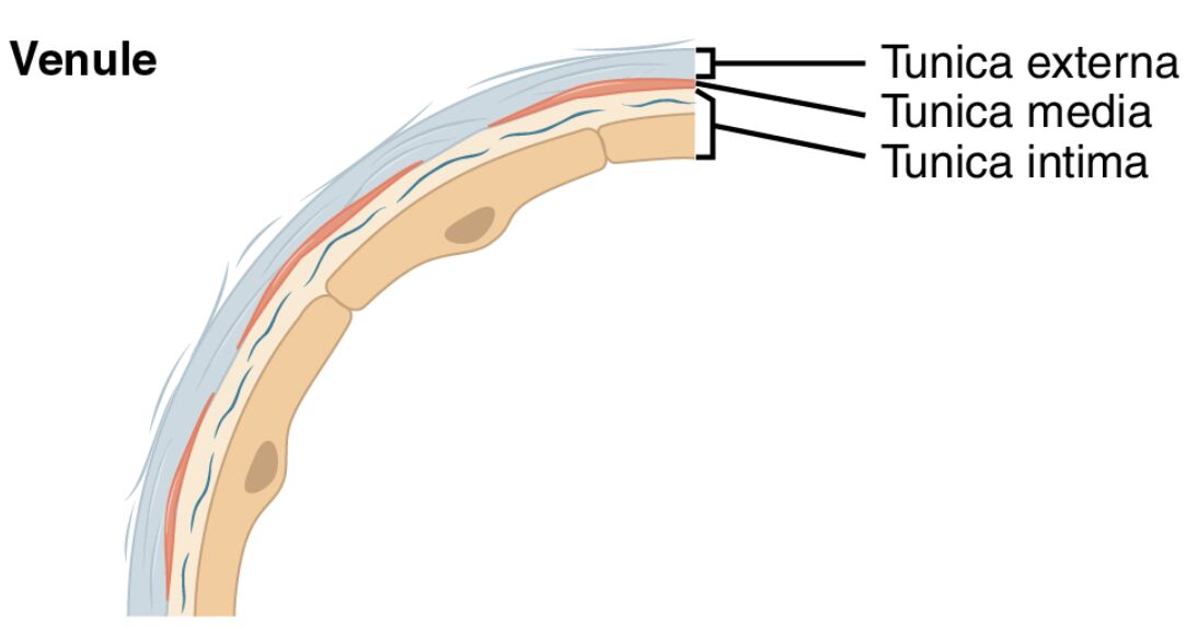
Tunica externa The tunica externa is the outermost layer of the venule, composed of a thin layer of connective tissue that provides minimal structural support. It helps anchor the venule to surrounding tissues and contains a few small vessels for nourishment.
Tunica media The tunica media is the middle layer, consisting of a sparse arrangement of smooth muscle cells and elastic fibers that offer limited contractile ability. This thin layer allows the venule to adjust slightly to changes in blood pressure and flow.
Tunica intima The tunica intima is the innermost layer, lined with a single layer of endothelial cells that reduce friction and support blood movement. Unlike larger veins, venules typically lack valves, relying on their connection to the venous system for directional flow.
Endothelial cell The endothelial cell forms the inner lining of the tunica intima, playing a key role in regulating blood flow and preventing clotting. These cells also facilitate the exchange of nutrients and gases as blood transitions from capillaries.
Lumen The lumen is the central cavity of the venule where blood flows, characterized by its small diameter measured in micrometers. This narrow space allows for the efficient collection of blood from capillary beds while maintaining low pressure.
The Role of Venules in the Circulatory System
Venules serve as a critical bridge between capillaries and veins in the circulatory network. Their structure supports the body’s ability to manage blood flow and nutrient exchange.
- The tunica externa provides a basic framework, anchoring the venule in tissue.
- The tunica media allows minor adjustments to accommodate blood volume changes.
- Endothelial cells facilitate the movement of hormones like T3 and T4 from the thyroid gland.
- This design ensures smooth transition of deoxygenated blood toward larger veins.
Anatomical Features of Venules Under the Microscope
The sectional view of a venule highlights its simplified yet functional anatomy. Each component contributes to its role in microcirculation.
- The lumen appears narrow, reflecting the venule’s role as a collector from capillaries.
- The tunica intima with endothelial cells forms a smooth inner surface.
- The tunica media is thin, indicating limited muscular control compared to larger veins.
- The tunica externa offers minimal support, suiting the venule’s small size.
Physiological Functions and Importance
The anatomy of venules enables them to perform essential physiological tasks. Their structure supports efficient blood collection and transport.
- The endothelial cell regulates the exchange of oxygen and carbon dioxide with tissues.
- The lumen collects blood at low pressure, preventing damage to delicate capillary networks.
- The tunica media assists in maintaining flow during minor pressure changes.
- This functionality is vital for tissue perfusion and waste removal.
Clinical Significance of Venule Anatomy
Understanding the structure of venules offers insights into potential health issues. Changes in these layers can indicate or contribute to circulatory challenges.
- Inflammation of endothelial cells can lead to increased permeability, causing edema.
- Damage to the tunica intima may contribute to microvascular complications in diabetes.
- The thin tunica media makes venules vulnerable to pressure changes in hypertension.
- Research into venule health supports treatments for vascular and inflammatory conditions.
Comparison with Other Blood Vessels
Venules differ from arteries, veins, and capillaries due to their unique microscopic features. This comparison highlights their specific role in the circulatory system.
- Unlike arteries, the tunica media in venules is much thinner, reflecting lower pressure.
- Larger veins feature valves, absent in venules due to their size and role.
- Capillaries have a single layer, while the tunica intima of venules adds slight thickness.
- The lumen of venules is smaller than that of veins, aligning with their transitional function.
Maintenance and Regulation of Venules
The body employs mechanisms to maintain the health and function of venules. These processes ensure optimal performance under varying conditions.
- Endothelial cells release nitric oxide to regulate local blood flow.
- The tunica externa is supported by minimal nourishment, suiting its thin structure.
- The tunica media responds to local metabolic signals to adjust flow.
- This regulation supports efficient transition of blood from capillaries to veins.
In conclusion, the sectional view of a venule, as depicted in this image, showcases a delicately balanced structure designed for efficient blood collection and transport. With its tunica externa, tunica media, tunica intima, endothelial cells, and lumen, this vessel exemplifies the circulatory system’s ability to adapt to the needs of microcirculation. Exploring these features deepens our understanding of venous anatomy and its critical role in sustaining tissue health.

