Large veins are vital components of the circulatory system, tasked with returning deoxygenated blood to the heart through a network of resilient and adaptable structures. This image offers a sectional view of a large vein, revealing its layered anatomy and the intricate elements that support its function in maintaining venous return.
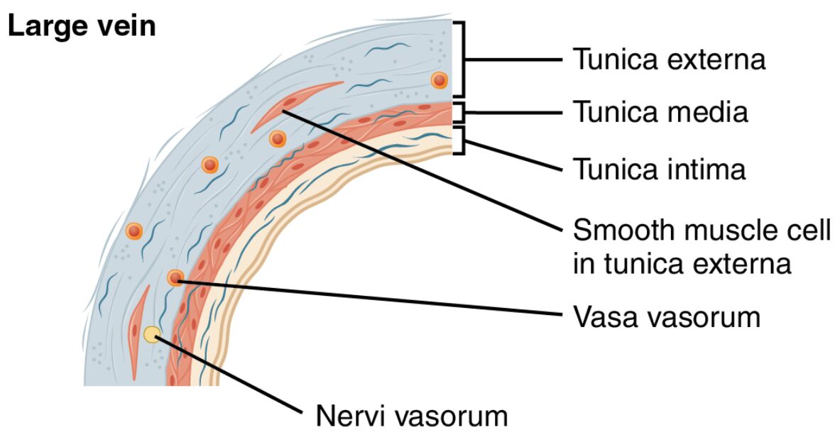
Tunica externa The tunica externa is the outermost layer of a large vein, made up of dense connective tissue that provides structural support and elasticity. It contains small blood vessels and nerves, ensuring the vein remains nourished and responsive to bodily needs.
Tunica media The tunica media is the middle layer, featuring a thin layer of smooth muscle cells and elastic fibers that aid in venous distensibility. This layer allows the vein to expand and accommodate varying blood volumes, particularly during periods of increased flow.
Tunica intima The tunica intima is the innermost layer, lined with endothelial cells that reduce friction and facilitate smooth blood flow. While it lacks valves in large veins, it plays a crucial role in preventing clot formation and supporting venous integrity.
Smooth muscle cell in tunica externa Smooth muscle cells in the tunica externa are sparsely distributed, contributing to the vein’s ability to adjust its diameter. These cells help maintain venous tone and assist in the return of blood against gravity.
Vasa vasorum The vasa vasorum are tiny blood vessels within the tunica externa that supply oxygen and nutrients to the vein wall. They are essential for the health of the thicker venous layers, particularly in larger veins where diffusion alone is insufficient.
Nervi vasorum The nervi vasorum are nerves embedded in the tunica externa that regulate vascular tone through autonomic signals. They enable the vein to respond to changes in blood pressure and support efficient circulation.
The Role of Large Veins in the Circulatory System
Large veins serve as the primary conduits for returning deoxygenated blood to the heart. Their robust structure supports the body’s ability to manage blood volume and pressure effectively.
- The tunica externa anchors the vein, providing stability and flexibility during movement.
- The tunica media allows veins to act as blood reservoirs, holding a significant portion of the body’s blood supply.
- Vasa vasorum ensure the vein wall remains healthy by delivering essential nutrients.
- This design supports venous return, especially in the lower extremities where gravity is a factor.
Anatomical Layers of Large Veins
The sectional view of a large vein reveals a complex arrangement of layers. Each layer contributes uniquely to the vein’s overall function and resilience.
- The tunica intima forms a smooth inner lining, minimizing resistance to blood flow.
- The tunica media contains elastic fibers that help the vein stretch and recoil.
- Smooth muscle cells in tunica externa provide additional support for venous tone.
- The nervi vasorum integrate neural control, enhancing vascular adaptability.
Physiological Functions and Importance
The anatomy of large veins enables them to perform critical physiological roles. Their structure ensures efficient blood transport and adaptation to bodily demands.
- The tunica media expands to accommodate increased venous return after physical activity.
- Vasa vasorum nourish the vein wall, preventing degeneration in larger vessels.
- The tunica intima facilitates the transport of hormones like T3 and T4 from the thyroid gland.
- This adaptability helps maintain circulation and prevent blood pooling.
Clinical Significance of Large Vein Anatomy
Understanding the structure of large veins offers insights into potential health issues. Changes in these layers can signal or contribute to circulatory disorders.
- Weakness in the tunica externa may lead to varicose veins, causing discomfort and swelling.
- Thickening of the tunica media can occur in chronic venous insufficiency, affecting blood flow.
- Damage to the tunica intima increases the risk of deep vein thrombosis.
- Research into vasa vasorum health supports treatments for vascular diseases.
Comparison with Other Vascular Structures
Large veins differ from arteries, capillaries, and smaller veins due to their specific design. This comparison highlights their unique role in the circulatory system.
- Unlike arteries, large veins have a thinner tunica media and rely less on muscular control.
- Capillaries lack the layered structure, focusing on exchange rather than transport.
- Smaller veins may include valves, absent in large veins due to their size and flow dynamics.
- The presence of nervi vasorum distinguishes large veins from less innervated vessels.
Maintenance and Regulation of Large Veins
The body employs mechanisms to maintain the health and function of large veins. These processes ensure optimal performance under varying physiological conditions.
- The nervi vasorum respond to sympathetic stimulation, adjusting venous tone.
- Smooth muscle cells in tunica externa contract to assist venous return during muscle activity.
- The vasa vasorum continuously supply nutrients, supporting the tunica externa integrity.
- Hormonal changes influence the tunica media to adapt to blood volume shifts.
In conclusion, the sectional view of a large vein, as depicted in this image, showcases a sophisticated vascular structure designed for efficient blood return. With its tunica externa, tunica media, tunica intima, smooth muscle cells in tunica externa, vasa vasorum, and nervi vasorum, this vein type exemplifies the body’s ability to manage circulation under diverse conditions. Exploring these features deepens our understanding of venous anatomy and its critical role in sustaining health.

