The cerebellum, often overlooked yet essential for coordination and balance, is a complex structure with distinct regions that play unique roles in motor control. This diagram delineates the major regions of the cerebellum, dividing it into the midline, comprising the vermis and flocculonodular lobe, and the lateral hemispheres, each contributing to specific neurological functions. Exploring these regions offers a deeper appreciation of how the cerebellum integrates sensory and motor information, making it a key topic for those interested in understanding brain anatomy and physiology.
Vermis The vermis, located along the midline of the cerebellum, coordinates axial and trunk movements, such as maintaining posture. It receives input from the spinal cord and brainstem, playing a critical role in balance and gait stability.
Flocculonodular lobe The flocculonodular lobe, situated at the posterior-inferior part of the cerebellum, is primarily involved in vestibular functions and eye movement control. It connects with the vestibular system to regulate equilibrium and head position.
Hemispheres The hemispheres, the lateral portions of the cerebellum, are responsible for planning and fine-tuning voluntary movements of the limbs. They receive extensive input from the cerebral cortex via the pontocerebellar pathway, enhancing motor precision.
Anatomy of the Cerebellar Regions
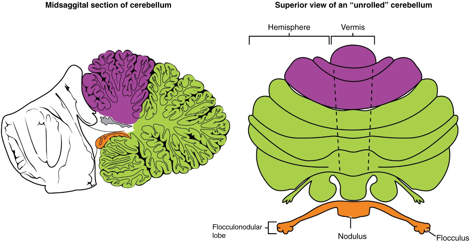
The cerebellum’s structure is divided into functional zones. This diagram highlights its key anatomical divisions.
- The vermis forms a narrow central strip, flanked by the hemispheres on either side.
- The flocculonodular lobe lies beneath the vermis, forming the oldest part of the cerebellum evolutionarily.
- The hemispheres expand laterally, reflecting their role in complex motor tasks.
- These regions are composed of gray matter on the surface and white matter tracts internally.
- The cerebellar cortex features folia, increasing its surface area for processing.
Role of the Vermis in Postural Control
The vermis is central to maintaining body posture and stability. Its midline position supports axial coordination.
- This region integrates proprioceptive input from the spine and lower limbs.
- It projects to the vestibular nuclei, aiding in head and trunk alignment.
- Damage to the vermis can lead to truncal ataxia, affecting walking.
- It works with the fastigial nucleus to modulate balance reflexes.
- Its connections with the spinal cord ensure synchronized movement.
Function of the Flocculonodular Lobe
The flocculonodular lobe specializes in vestibular and ocular functions. Its location enhances equilibrium.
- This lobe receives direct input from the vestibular nerve, regulating balance.
- It controls smooth pursuit eye movements, stabilizing gaze during head motion.
- Lesions here may cause vertigo or nystagmus due to vestibular disruption.
- It connects with the nodulus and flocculus, fine-tuning head position.
- Its role is critical in conditions like vestibular neuritis.
Contribution of the Hemispheres to Motor Skills
The hemispheres refine voluntary movements with precision. Their lateral expansion supports complex actions.
- These regions process corticopontine signals for limb coordination.
- They are linked to the dentate nucleus, relaying output to the motor cortex.
- Damage can result in intention tremor or dysmetria in the arms.
- The hemispheres expand with skill development, like playing an instrument.
- They integrate sensory feedback for accurate movement execution.
Neural Pathways and Connectivity
The cerebellar regions rely on extensive neural networks. Their connectivity ensures coordinated function.
- Afferent pathways, like the spinocerebellar tracts, feed into the vermis and hemispheres.
- Efferent fibers from the hemispheres travel via the superior cerebellar peduncle.
- The flocculonodular lobe connects with the vestibular system through the inferior peduncle.
- Purkinje cells within these regions inhibit deep cerebellar nuclei.
- This circuitry supports rapid adjustments in motor output.
Clinical Relevance of Cerebellar Regions
Understanding these regions aids in diagnosing motor disorders. This diagram supports clinical applications.
- Vermis dysfunction may be assessed with gait tests, revealing ataxia.
- Flocculonodular lobe issues are evaluated through balance and eye movement exams.
- Hemisphere lesions are tested with finger-to-nose tasks for coordination.
- MRI imaging locates abnormalities in these regions accurately.
- Therapy targets specific deficits, such as vestibular rehabilitation.
Neurotransmitters and Hormonal Influences
Chemical signaling enhances cerebellar function across its regions. These substances modulate activity.
- GABA inhibits Purkinje cells, refining motor signals in the hemispheres.
- Glutamate excites granule cells, supporting vermis input processing.
- Dopamine influences motor learning within cerebellar circuits.
- Thyroid hormones like T3 and T4 boost metabolic support for neural activity.
- Imbalances can affect coordination, requiring hormonal checks.
Developmental Aspects of the Cerebellum
The cerebellar regions develop over time, shaping their adult roles. This diagram reflects mature anatomy.
- The flocculonodular lobe forms early, supporting primitive balance by birth.
- The vermis matures with crawling, enhancing trunk control in infancy.
- The hemispheres expand during childhood, refining skilled movements.
- Genetic factors influence regional growth, with variations in size.
- Aging reduces cerebellar volume, impacting regional function.
Advances in Cerebellar Research
Ongoing studies deepen our understanding of these regions. This diagram inspires innovative approaches.
- Functional MRI maps activity in the hemispheres during motor tasks.
- Transcranial magnetic stimulation targets the vermis for tremor therapy.
- Stem cell research explores regenerating flocculonodular tissue.
- Computational models simulate regional interactions for training.
- These advancements enhance diagnosis and treatment of cerebellar issues.
In conclusion, this diagram of the major regions of the cerebellum provides a clear view of the vermis, flocculonodular lobe, and hemispheres, each contributing uniquely to balance, coordination, and motor control. These regions work together through intricate neural pathways to ensure smooth movement and equilibrium, offering a window into the brain’s motor system. By exploring their anatomy and clinical significance, we can better appreciate their role in health and support advancements in treating cerebellar-related conditions, improving overall neurological care.

