The cochlea, a spiral structure within the inner ear, relies on its hair cells to convert sound vibrations into electrical signals, a process beautifully illustrated in this image. This image highlights the scala tympani, scala vestibuli, cochlear duct, and the organ of Corti, where mechanoreceptor hair cells reside atop the basilar membrane, playing a central role in hearing. This article provides a detailed examination of these components, exploring their anatomical layout and physiological significance in the auditory system.
Labeled Parts of the Cochlea
Scala tympani The scala tympani is a lower fluid-filled chamber of the cochlea that extends from the apex to the round window, dissipating pressure waves generated by sound. It maintains fluid equilibrium, ensuring the cochlea can process auditory signals without structural damage.
Scala vestibuli The scala vestibuli is an upper fluid-filled chamber that receives pressure waves from the oval window, transmitting them toward the cochlear apex. It collaborates with the scala tympani to facilitate fluid movement that stimulates hair cells for sound detection.
Cochlear duct The cochlear duct, or scala media, is a central chamber between the scala vestibuli and scala tympani, filled with endolymph and housing the organ of Corti. It serves as the sensory core of the cochlea, separating the perilymphatic scalae with its specialized membranes.
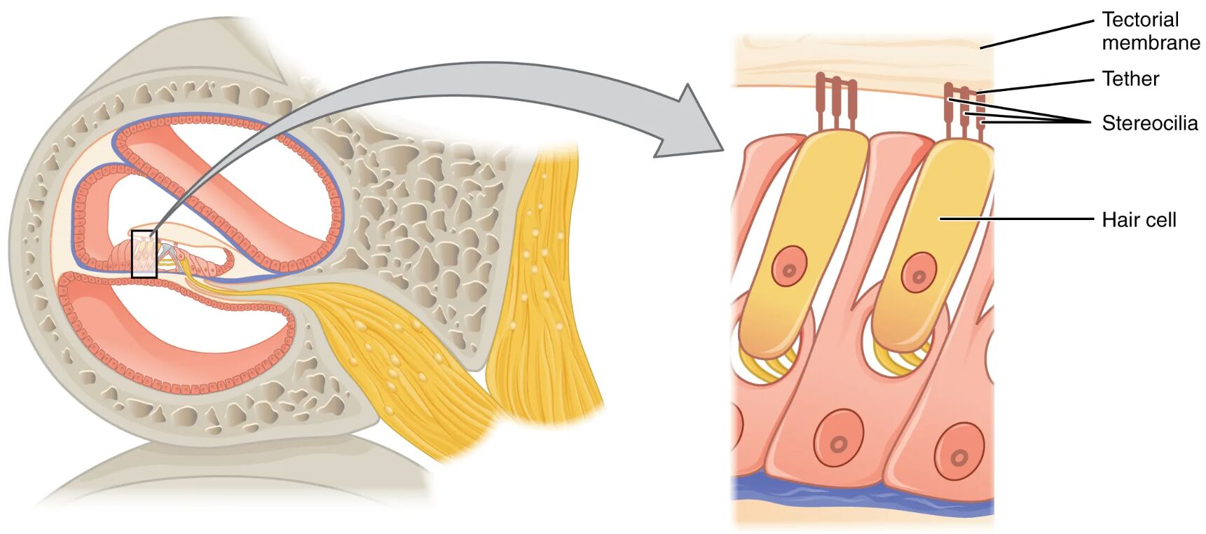
Organ of Corti The organ of Corti is a structure within the cochlear duct, resting on the basilar membrane, and contains hair cells that detect sound vibrations. It converts mechanical energy into electrical signals, transmitting them to the auditory nerve for hearing perception.
Basilar membrane The basilar membrane supports the organ of Corti and varies in width and stiffness along the cochlea, tuning it to different sound frequencies. Its movement in response to fluid pressure waves is key to activating specific hair cells for pitch discrimination.
Hair cells Hair cells are mechanoreceptor cells within the organ of Corti, featuring stereocilia that bend with sound-induced fluid motion to generate electrical impulses. They are essential for transducing auditory signals to the cochlear nerve, enabling the brain to interpret sound.
Anatomical Overview of the Cochlea
The cochlea’s cross-sectional design reveals a sophisticated interplay of fluid-filled spaces and sensory structures, optimized for auditory processing. This arrangement ensures that sound vibrations are efficiently transformed into neural signals.
- Fluid compartments: The scala tympani and scala vestibuli encase the cochlear duct, creating a dual perilymph system that supports pressure wave propagation.
- Sensory location: The organ of Corti, embedded in the cochlear duct, is the site of auditory transduction, anchored by the basilar membrane.
- Fluid separation: The cochlear duct’s endolymph contrasts with the perilymph in the scalae, providing the ionic environment needed for hair cell function.
- Membrane gradient: The basilar membrane’s varying properties allow it to resonate at specific frequencies, enhancing the cochlea’s frequency resolution.
- Cellular precision: Hair cells are strategically aligned to detect fluid movements, forming the critical link between mechanical and electrical signals.
Physiological Functions of Hair Cells
Hair cells within the cochlea are the linchpin of auditory transduction, converting mechanical vibrations into electrical impulses for sound perception. Their physiological roles are finely tuned to handle diverse auditory inputs.
- Vibration detection: Hair cells’ stereocilia bend in response to pressure waves in the cochlear fluid, initiated by sound vibrations.
- Signal generation: This bending opens ion channels, depolarizing the hair cells and releasing neurotransmitters to activate auditory nerve fibers.
- Frequency tuning: The basilar membrane’s resonance at different points along the cochlea allows hair cells to detect specific sound frequencies.
- Amplification: Outer hair cells actively amplify sound through electromotility, enhancing the sensitivity of the inner hair cells.
- Neural transmission: Inner hair cells primarily transmit signals to the brain, ensuring accurate representation of sound pitch and intensity.
Developmental and Cellular Dynamics
The cochlea, including its hair cells, develops during embryogenesis, maturing to support hearing by early childhood. This process involves coordinated cellular growth and differentiation essential for auditory function.
- Embryonic origins: The cochlear duct forms from the otic vesicle, with the scalae developing as perilymphatic spaces around it.
- Hair cell formation: Hair cells differentiate within the organ of Corti, with stereocilia aligning to detect fluid motion during fetal development.
- Membrane development: The basilar membrane grows with a gradient of stiffness, enabling frequency-specific responses as the cochlea spirals.
- Fluid establishment: Perilymph and endolymph fill the scalae and cochlear duct, respectively, creating the environment for sound transduction.
- Postnatal maturation: Neural connections with hair cells strengthen after birth, refining the cochlea’s sensitivity to sound.
Clinical Relevance and Cochlear Health
Understanding the role of hair cells in the cochlea is crucial for diagnosing and managing hearing-related conditions. Clinical assessments often focus on these cells to identify auditory impairments.
- Sensorineural hearing loss: Damage to hair cells, often from prolonged noise exposure or ototoxic drugs, impairs sound transduction in the organ of Corti.
- Presbycusis: Age-related degeneration of hair cells and the basilar membrane leads to high-frequency hearing loss.
- Tinnitus: Persistent ringing may result from hair cell dysfunction or cochlear damage, affecting quality of life.
- Diagnostic tools: Otoacoustic emission tests and cochlear imaging evaluate hair cell function and structural integrity.
- Therapeutic options: Cochlear implants bypass damaged hair cells, while protective measures like noise reduction aim to preserve remaining cells.
In conclusion, the hair cells in the cochlea, as depicted in this cross-sectional image, are the heart of auditory perception, nestled within the organ of Corti atop the basilar membrane. The interplay of the scala tympani, scala vestibuli, and cochlear duct supports their function, transforming sound vibrations into the rich auditory experiences we enjoy. Exploring these structures deepens our understanding of hearing mechanisms and highlights the importance of protecting cochlear health for long-term auditory well-being.

