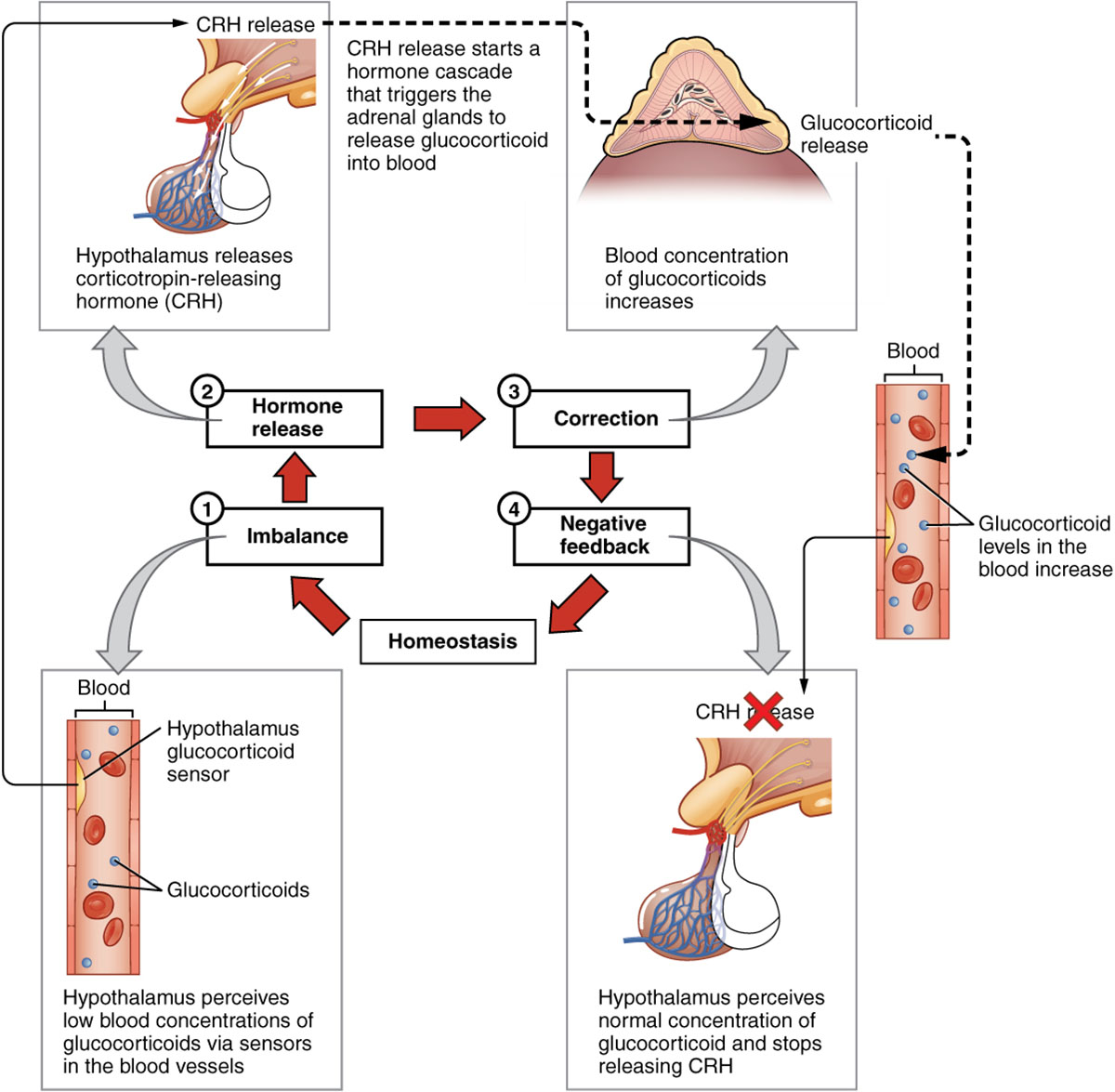
The body maintains balance through intricate feedback mechanisms, with the negative feedback loop playing a central role in regulating hormone levels and preventing overproduction. This diagram illustrates how the release of adrenal glucocorticoids is stimulated by hormones from the hypothalamus and pituitary gland, and how elevated glucocorticoid levels trigger negative signals to inhibit further hormone release from these glands. Exploring this image provides a clear insight into the dynamic process that ensures hormonal homeostasis.

Labelled Parts Explanation
- Adrenal glucocorticoids The adrenal glucocorticoids, such as cortisol, are steroid hormones produced by the adrenal cortex to manage stress and regulate metabolism. Their release is controlled by upstream hormonal signals but inhibited when levels rise too high, maintaining balance.
- Hypothalamus The hypothalamus, located in the brain, releases corticotropin-releasing hormone (CRH) to stimulate the pituitary gland, initiating the stress response. It receives negative feedback from elevated adrenal glucocorticoids to reduce CRH secretion.
- Pituitary gland The pituitary gland, situated at the brain’s base, secretes adrenocorticotropic hormone (ACTH) in response to CRH, prompting the adrenal glands to produce adrenal glucocorticoids. It is inhibited by high glucocorticoid levels, halting ACTH release via negative feedback.
- Negative signals The negative signals are feedback mechanisms triggered by elevated adrenal glucocorticoids levels, sending inhibitory messages to the hypothalamus and pituitary gland. This reduces CRH and ACTH production, preventing excessive hormone release and maintaining equilibrium.
Anatomical Overview of the Negative Feedback Loop
The negative feedback loop is a critical regulatory mechanism within the endocrine system, ensuring hormone levels remain within a healthy range. This diagram focuses on the interaction between the hypothalamus, pituitary gland, adrenal glucocorticoids, and negative signals in the hypothalamic-pituitary-adrenal (HPA) axis.
- The hypothalamus initiates the cascade by releasing CRH.
- The pituitary gland responds with ACTH, stimulating the adrenal glands.
- The adrenal glucocorticoids rise to address stress or metabolic needs.
- The negative signals feed back to suppress further hormone production.
This loop exemplifies endocrine self-regulation.
Role of the Hypothalamus in Feedback
The hypothalamus serves as the starting point of the feedback loop. Its function drives the stress response.
- The hypothalamus releases CRH to activate the pituitary gland.
- This hormone stimulates ACTH production under stress conditions.
- Elevated adrenal glucocorticoids inhibit CRH via negative signals.
- The process adjusts to maintain stress hormone levels.
This regulation is key to stress management.
Function of the Pituitary Gland
The pituitary gland acts as a mediator in the feedback loop. Its role connects hypothalamic and adrenal activity.
- The pituitary gland secretes ACTH in response to CRH.
- This hormone triggers adrenal glucocorticoids release from the adrenal cortex.
- High glucocorticoid levels send negative signals to reduce ACTH.
- The gland fine-tunes the stress response.
This step ensures balanced hormone output.
Significance of Adrenal Glucocorticoids
Adrenal glucocorticoids are the end product of the feedback loop. Their levels dictate the response.
- The adrenal glucocorticoids like cortisol regulate metabolism and immune response.
- They are produced in response to ACTH stimulation.
- Elevated levels initiate negative signals to the hypothalamus and pituitary gland.
- This maintains a stable internal environment.
This hormone is crucial for stress adaptation.
Mechanism of Negative Signals
Negative signals provide the corrective action in the loop. Their role prevents overproduction.
- The negative signals are triggered by high adrenal glucocorticoids levels.
- They inhibit CRH release from the hypothalamus and ACTH from the pituitary gland.
- This feedback reduces adrenal stimulation, lowering glucocorticoid output.
- The process operates continuously to adjust hormone levels.
This mechanism ensures homeostasis.
Physiological Importance of the Feedback Loop
The negative feedback loop maintains hormonal balance essential for health. Its design supports adaptive responses.
- The hypothalamus and pituitary gland initiate and modulate the stress response.
- The adrenal glucocorticoids address immediate and chronic stress.
- The negative signals prevent excessive glucocorticoid production.
- This regulation protects against stress-related damage.
The loop is vital for physiological stability.
Clinical Relevance of the Negative Feedback Loop
Understanding the negative feedback loop aids in diagnosing endocrine disorders. These components are key clinical markers.
- Dysfunction in the hypothalamus can lead to adrenal insufficiency, causing fatigue.
- Overactive pituitary gland activity may result in Cushing’s disease with high adrenal glucocorticoids.
- Impaired negative signals can cause chronic stress or hypercortisolism.
- Hormone levels are monitored via blood tests for treatment.
This knowledge guides endocrine therapy.
Conclusion
The negative feedback loop diagram provides a detailed view of how adrenal glucocorticoids, hypothalamus, pituitary gland, and negative signals work together to regulate hormone levels within the HPA axis. By exploring the stimulation and inhibition processes that maintain glucocorticoid balance, one gains insight into the body’s self-regulating mechanisms. This understanding serves as a foundation for studying endocrinology and addressing related health concerns, encouraging further exploration of the intricate feedback systems that sustain hormonal homeostasis and overall well-being.

