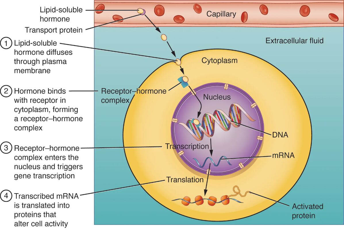
Lipid-soluble hormones, such as steroid hormones, play a crucial role in regulating gene expression and protein production within target cells, distinguishing them from water-soluble hormones. This diagram illustrates the process where a steroid hormone diffuses through the cell membrane, binds to a receptor in the cytosol, forms a receptor–hormone complex, enters the nucleus, binds to a target gene on DNA, and initiates messenger RNA (mRNA) and protein synthesis in the cytoplasm. Exploring this image provides a comprehensive understanding of how these hormones exert their effects at the cellular level.
Labelled Parts Explanation
- Steroid hormone The steroid hormone, derived from cholesterol, is a lipid-soluble molecule that easily crosses the cell membrane to enter target cells. It binds to specific receptors to initiate gene expression, influencing processes like metabolism and reproduction.
- Receptor in the cytosol The receptor in the cytosol is a protein that resides within the cell’s cytoplasm, awaiting binding with a steroid hormone. Upon binding, it undergoes a conformational change, enabling it to act as a transcription factor.
- Receptor–hormone complex The receptor–hormone complex forms when the steroid hormone binds to its cytosolic receptor, activating the complex for nuclear entry. This complex then translocates to the nucleus to regulate gene activity.
- Nucleus The nucleus houses the cell’s DNA and serves as the site where the receptor–hormone complex binds to target genes. It is the control center for protein synthesis, directing genetic expression based on hormonal signals.
- Target gene on DNA The target gene on DNA is a specific DNA sequence that the receptor–hormone complex binds to, initiating transcription. This binding activates the production of specific proteins tailored to the hormone’s function.
- Messenger RNA (mRNA) The messenger RNA (mRNA) is synthesized from the target gene through transcription, carrying the genetic code from the nucleus to the cytoplasm. It serves as a template for protein synthesis, translating the hormone’s signal into action.
- Protein synthesis The protein synthesis process occurs in the cytoplasm, where mRNA is translated by ribosomes to produce proteins that carry out the hormone’s effects. This step completes the hormone’s influence on cellular function.
- Cytoplasm The cytoplasm is the cellular compartment where protein synthesis takes place, containing ribosomes and other machinery for translation. It is the site where the final products of hormonal signaling are assembled.
Anatomical Overview of Lipid-Soluble Hormone Action
Lipid-soluble hormones, primarily steroids, exert their effects by directly influencing gene expression within target cells. This diagram outlines the step-by-step process of how these hormones interact with cellular components.
- The steroid hormone enters the cell due to its lipid solubility.
- The receptor in the cytosol and receptor–hormone complex facilitate nuclear entry.
- The nucleus and target gene on DNA drive transcriptional activity.
- The messenger RNA (mRNA), protein synthesis, and cytoplasm complete the protein production cycle.
This mechanism highlights the hormone’s intracellular pathway.
Role of the Steroid Hormone
The steroid hormone initiates the entire process with its unique properties. Its entry is the first critical step.
- The steroid hormone diffuses through the lipid bilayer of the cell membrane.
- It is produced by glands like the adrenal cortex or gonads.
- This diffusion allows direct access to intracellular targets.
- Examples include cortisol and testosterone.
This step sets the stage for gene regulation.
Function of the Receptor and Complex Formation
The receptor and its complex are pivotal for hormone action. Their interaction drives nuclear effects.
- The receptor in the cytosol binds the steroid hormone with high specificity.
- The receptor–hormone complex activates upon binding, changing its shape.
- This complex is transported into the nucleus for gene interaction.
- The process ensures targeted hormonal response.
This binding is essential for signal transduction.
Significance of the Nucleus and Target Gene
The nucleus and target gene are the sites of genetic activation. Their role is central to protein production.
- The nucleus protects DNA and facilitates transcription.
- The target gene on DNA is selectively activated by the receptor–hormone complex.
- This binding promotes mRNA synthesis for specific proteins.
- The process is highly regulated to match cellular needs.
This step translates hormonal signals into action.
Process of mRNA and Protein Synthesis
mRNA and protein synthesis are the outcomes of hormonal signaling. Their production fulfills the hormone’s purpose.
- The messenger RNA (mRNA) carries the genetic code to the cytoplasm.
- The protein synthesis occurs at ribosomes, building functional proteins.
- This process is guided by the hormone’s initial signal.
- The resulting proteins regulate cellular activities.
This translation is critical for physiological effects.
Physiological Importance of Lipid-Soluble Hormone Binding
The binding mechanism of lipid-soluble hormones ensures precise regulation. Its efficiency supports homeostasis.
- The steroid hormone’s ability to enter cells allows direct gene control.
- The receptor–hormone complex targets specific genes for transcription.
- The messenger RNA (mRNA) and protein synthesis produce long-term effects.
- This pathway adapts to metabolic or developmental needs.
The process is vital for sustained regulation.
Clinical Relevance of Hormone Binding
Understanding lipid-soluble hormone binding aids in diagnosing endocrine disorders. These components are key clinical markers.
- Excess steroid hormone activity can lead to Cushing’s syndrome, causing weight gain.
- Deficient receptor in the cytosol function may result in hormone resistance.
- Abnormal protein synthesis can indicate genetic or hormonal imbalances.
- Hormone levels are monitored via blood tests for treatment.
This knowledge guides endocrine management.
Conclusion
The binding of lipid-soluble hormones diagram provides a detailed view of the process involving the steroid hormone, receptor in the cytosol, receptor–hormone complex, nucleus, target gene on DNA, messenger RNA (mRNA), protein synthesis, and cytoplasm, illustrating how these hormones regulate gene expression. By exploring the mechanism of diffusion, receptor binding, and protein production, one gains insight into the intricate cellular responses driven by these hormones. This understanding serves as a foundation for studying endocrinology and addressing related health concerns, encouraging further exploration of the sophisticated interplay between lipid-soluble hormones and cellular function.

