The heart’s ability to beat rhythmically depends on its specialized conduction system, a network that coordinates electrical impulses for efficient pumping. This anterior view of a frontal section diagram illustrates key components such as the sinoatrial node, internodal pathways, atrioventricular node, atrioventricular bundle, right bundle branch, left bundle branch, and Purkinje fibers, offering a clear view of how these structures regulate cardiac activity. Exploring this image provides a deeper understanding of the electrical framework that sustains circulation.
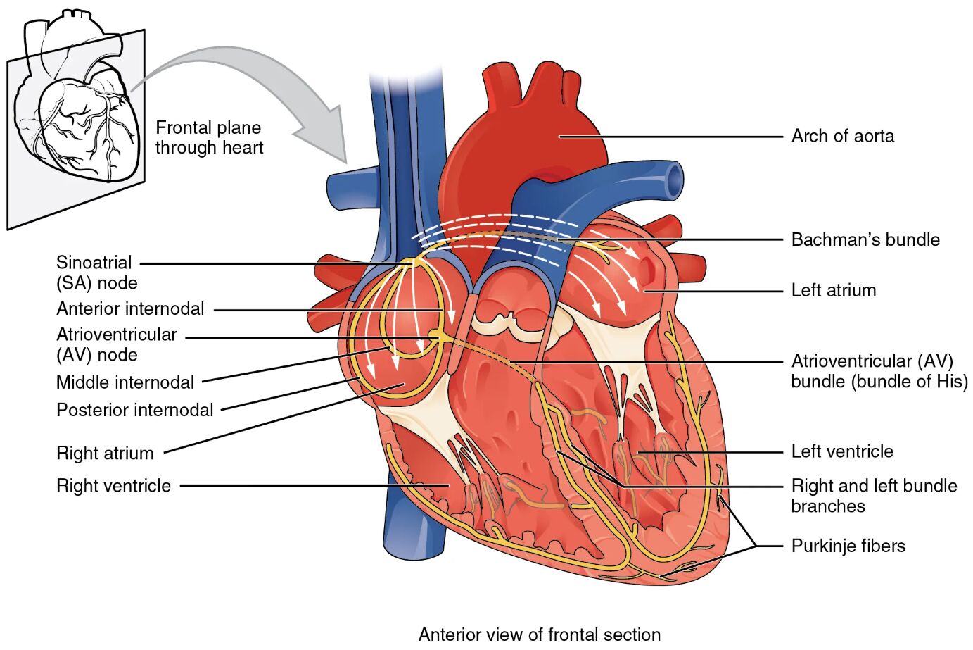
Labelled Parts Explanation
- Sinoatrial node The sinoatrial node, located in the right atrium, serves as the heart’s natural pacemaker, initiating electrical impulses. It sets the rhythm for atrial contraction and influences the overall heart rate.
- Internodal pathways The internodal pathways are specialized tracts within the atria that conduct impulses from the sinoatrial node to the atrioventricular node. They ensure rapid and coordinated atrial depolarization.
- Atrioventricular node The atrioventricular node, situated near the atrioventricular septum, delays the electrical impulse to allow atrial contraction before ventricular activation. This delay is crucial for effective ventricular filling.
- Atrioventricular bundle The atrioventricular bundle, or bundle of His, transmits the electrical impulse from the atrioventricular node to the bundle branches. It serves as the sole electrical connection between the atria and ventricles.
- Right bundle branch The right bundle branch carries the impulse down the right side of the interventricular septum to the right ventricle. It ensures synchronized contraction of the right ventricular muscle.
- Left bundle branch The left bundle branch conducts the impulse to the left ventricle, dividing into anterior and posterior fascicles. It coordinates the left ventricular contraction for systemic circulation.
- Purkinje fibers The Purkinje fibers are specialized fibers that distribute the impulse rapidly across the ventricular myocardium. They enable the ventricles to contract almost simultaneously for efficient pumping.
Anatomical Overview of the Cardiac Conduction System
The cardiac conduction system is a sophisticated network that regulates the heart’s rhythm. This frontal section view highlights the pathway that ensures coordinated contractions.
- The sinoatrial node initiates the electrical signal, starting the cardiac cycle.
- The internodal pathways and atrioventricular node relay and delay the impulse for atrial and ventricular synchronization.
- The atrioventricular bundle and right bundle branch along with left bundle branch distribute the signal to the ventricles.
- The Purkinje fibers finalize the process, triggering ventricular contraction.
This system maintains the heart’s ability to pump blood effectively.
Role of the Sinoatrial Node and Internodal Pathways
The sinoatrial node and internodal pathways kickstart the cardiac cycle. Their function is vital for rhythm initiation.
- The sinoatrial node generates impulses at a rate of 60-100 beats per minute under normal conditions.
- The internodal pathways ensure the signal spreads quickly across the atria for unified contraction.
- These pathways include the anterior, middle, and posterior internodal tracts.
- The process sets the stage for atrial systole before ventricular action.
This initial step is essential for heart rate regulation.
Function of the Atrioventricular Node and Bundle
The atrioventricular node and bundle manage the transition from atria to ventricles. Their role ensures proper timing.
- The atrioventricular node delays the impulse by about 0.1 seconds, allowing atrial emptying.
- The atrioventricular bundle then carries the signal through the interventricular septum.
- This delay prevents atrial and ventricular contractions from overlapping.
- The bundle’s location makes it a critical point for electrical conduction.
This mechanism supports efficient blood flow coordination.
Significance of Bundle Branches and Purkinje Fibers
The bundle branches and Purkinje fibers complete the conduction process. They ensure ventricular synchronization.
- The right bundle branch and left bundle branch distribute the impulse to their respective ventricles.
- The Purkinje fibers spread the signal rapidly through the ventricular walls.
- Their large diameter and fast conduction velocity maximize contraction efficiency.
- This network allows the ventricles to eject blood almost simultaneously.
These components are key to maintaining cardiac output.
Physiological Importance of the Conduction System
The conduction system’s design optimizes the heart’s pumping action. Its structure supports continuous circulation.
- The sinoatrial node’s pacemaker activity adapts to physiological demands via autonomic input.
- The internodal pathways and atrioventricular node ensure a controlled sequence of events.
- The Purkinje fibers’ rapid conduction enhances ventricular ejection force.
- This system maintains a consistent heart rate and rhythm.
The coordination is vital for cardiovascular stability.
Clinical Relevance of the Conduction System
Understanding the conduction system aids in diagnosing and treating cardiac arrhythmias. These components are key clinical targets.
- Dysfunction of the sinoatrial node can lead to sick sinus syndrome, causing irregular heart rates.
- Blockage of the atrioventricular node or atrioventricular bundle results in heart block.
- Damage to the right bundle branch or left bundle branch may cause bundle branch block.
- The Purkinje fibers’ impairment can lead to ventricular fibrillation, a life-threatening condition.
This knowledge guides interventions like pacemakers and ablation therapies.
Conclusion
The conduction system of the heart, as depicted in this anterior view of a frontal section, provides a detailed look at the electrical network that drives cardiac rhythm. By exploring the sinoatrial node, internodal pathways, atrioventricular node, atrioventricular bundle, right bundle branch, left bundle branch, and Purkinje fibers, one gains insight into how these specialized structures coordinate the heart’s contractions. This understanding serves as a foundation for studying cardiovascular physiology and addressing related health issues, encouraging further exploration of the heart’s intricate electrical design and its critical role in sustaining life.

