The human body relies on a sophisticated circulatory system to deliver oxygen and nutrients while removing waste products. This diagram provides a comprehensive view of blood circulation, illustrating the flow between the heart, lungs, and body tissues through a network of arteries, veins, and capillaries. Exploring this image offers valuable insights into the physiological processes that sustain life and maintain homeostasis.
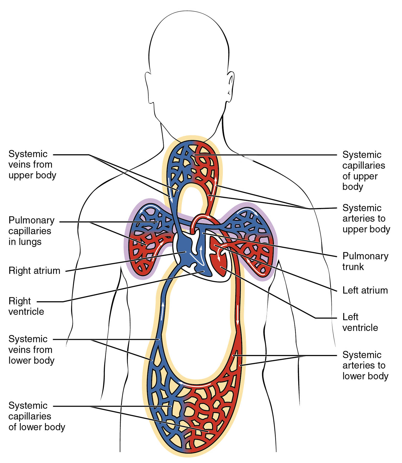
Labelled Parts Explanation
- Systemic veins from upper body The systemic veins from the upper body collect deoxygenated blood from the head, neck, and arms. This blood is transported back to the heart for reoxygenation.
- Pulmonary capillaries in lungs The pulmonary capillaries in the lungs facilitate the exchange of oxygen and carbon dioxide between the air and blood. Oxygenated blood then returns to the heart via the pulmonary veins.
- Right atrium The right atrium receives deoxygenated blood from the systemic veins, including those from the upper and lower body. It contracts to push blood into the right ventricle.
- Right ventricle The right ventricle pumps deoxygenated blood into the pulmonary trunk for delivery to the lungs. Its muscular structure supports the lower pressure of pulmonary circulation.
- Systemic veins from lower body The systemic veins from the lower body gather deoxygenated blood from the legs and abdomen. This blood is returned to the right atrium to complete the systemic cycle.
- Systemic capillaries of lower body The systemic capillaries of the lower body enable the exchange of nutrients, oxygen, and waste products with tissues. Deoxygenated blood is then collected by veins for return to the heart.
- Systemic capillaries of upper body The systemic capillaries of the upper body support metabolic exchanges in the head, neck, and arms. They ensure oxygen delivery and carbon dioxide removal at the cellular level.
- Systemic arteries to upper body The systemic arteries to the upper body distribute oxygenated blood from the aorta to the head, neck, and arms. These arteries branch into smaller vessels to reach tissues.
- Pulmonary trunk The pulmonary trunk carries deoxygenated blood from the right ventricle to the lungs. It divides into the pulmonary arteries to facilitate gas exchange.
- Left atrium The left atrium receives oxygenated blood from the pulmonary veins after gas exchange in the lungs. It contracts to send blood into the left ventricle.
- Left ventricle The left ventricle pumps oxygenated blood into the aorta for systemic circulation. Its thick walls generate the high pressure needed to supply the entire body.
- Systemic arteries to lower body The systemic arteries to the lower body deliver oxygenated blood from the aorta to the legs and abdomen. They ensure adequate perfusion of lower body tissues.
Anatomical Overview of Blood Circulation
The circulatory system is a vital network that sustains the body’s functions. This diagram highlights the dual pathways of pulmonary and systemic circulation, essential for oxygen delivery and waste elimination.
- The pulmonary capillaries in lungs and pulmonary trunk form the pulmonary circuit, where blood is oxygenated.
- The right atrium and right ventricle initiate this process by receiving and pumping deoxygenated blood.
- The left atrium and left ventricle drive systemic circulation, distributing oxygenated blood via the systemic arteries to upper body and systemic arteries to lower body.
- Systemic veins from upper body and systemic veins from lower body return deoxygenated blood to the heart, completing the cycle.
This intricate system ensures that every tissue receives the necessary oxygen and nutrients while removing metabolic byproducts.
Pulmonary Circulation Process
Pulmonary circulation is the first step in refreshing the blood’s oxygen supply. It involves the movement of blood from the heart to the lungs and back.
- The right ventricle pumps blood into the pulmonary trunk, which splits to reach the lungs.
- Within the pulmonary capillaries in lungs, oxygen from inhaled air diffuses into the blood, while carbon dioxide is expelled.
- Oxygenated blood then flows back to the left atrium via the pulmonary veins (not labeled but implied).
- This process is critical for maintaining adequate oxygen levels in the bloodstream.
Efficient pulmonary circulation supports overall respiratory function and cardiovascular health.
Systemic Circulation Explained
Systemic circulation delivers oxygenated blood to the body’s tissues and returns deoxygenated blood to the heart. This pathway is powered by the left side of the heart.
- The left ventricle ejects blood into the systemic arteries to upper body and systemic arteries to lower body.
- Systemic capillaries of upper body and systemic capillaries of lower body facilitate the exchange of oxygen, nutrients, and waste with cells.
- Deoxygenated blood is collected by the systemic veins from upper body and systemic veins from lower body, returning it to the right atrium.
- This circuit ensures that all organs and tissues receive the resources needed for function.
The systemic circuit’s high pressure, driven by the left ventricle, is essential for effective distribution.
Role of Capillaries in Circulation
Capillaries are the smallest blood vessels, playing a pivotal role in exchange processes. They connect arteries and veins, enabling critical interactions.
- The pulmonary capillaries in lungs allow gas exchange, enriching blood with oxygen.
- Systemic capillaries of upper body and systemic capillaries of lower body deliver oxygen and nutrients while removing carbon dioxide and waste.
- Their thin walls enhance diffusion, making them ideal for metabolic exchanges.
- Capillary networks adapt to local tissue needs, ensuring efficient circulation.
This microcirculation is fundamental to maintaining cellular health and homeostasis.
Physiological Importance of Heart Chambers
The heart’s chambers are designed to handle specific circulatory tasks. Their structure supports the demands of both pulmonary and systemic circuits.
- The right atrium and left atrium serve as entry points for deoxygenated and oxygenated blood, respectively.
- The right ventricle and left ventricle act as pumps, with the left side generating higher pressure for systemic flow.
- The left ventricle’s thicker myocardium reflects its role in systemic circulation, while the right ventricle is adapted for pulmonary pressure.
- Coordinated chamber function ensures continuous blood flow throughout the body.
This anatomical design optimizes the heart’s efficiency in sustaining life.
Conclusion
The diagram of blood circulation in the human body provides a clear roadmap of this life-sustaining process. By understanding the roles of the heart chambers, arteries, veins, and capillaries, one gains insight into how oxygen and nutrients are delivered while waste is removed. This knowledge lays a strong foundation for exploring cardiovascular physiology and its clinical applications, fostering a deeper appreciation for the body’s intricate design. Regular study of such diagrams enhances comprehension of circulatory dynamics, encouraging further exploration of related topics.

