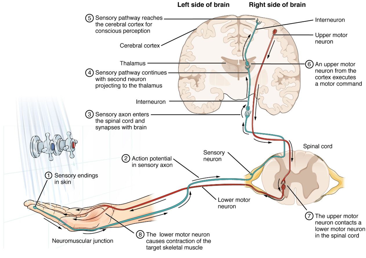
The nervous system orchestrates a seamless flow of information from sensory detection to motor execution, enabling responses to environmental stimuli like water temperature on the skin. This illustrative diagram traces the pathway from peripheral sensory endings through the spinal cord and brain to muscle contraction, highlighting the roles of sensory neurons, interneurons, and motor neurons in both ascending sensory and descending motor tracts. Such integration allows for conscious perception in the cerebral cortex and precise motor commands, essential for adaptive behaviors and maintaining homeostasis in daily activities.

Labeled Steps in the Pathway
Sensory endings in skin
The sensory endings in skin are specialized receptors, such as thermoreceptors, that detect stimuli like temperature changes and generate graded potentials proportional to stimulus intensity. These endings initiate the sensory pathway by transducing physical signals into electrical ones, ensuring accurate environmental monitoring.
Action potential in sensory axon
The action potential in sensory axon propagates along the peripheral process of the sensory neuron if the graded potential reaches threshold at the initial segment. This all-or-none signal travels rapidly to the spinal cord, preserving stimulus information through frequency coding.
Sensory axon enters the spinal cord and synapses with brain
The sensory axon enters the spinal cord and synapses with brain via connections in the dorsal horn, where it releases neurotransmitters to excite second-order neurons. This synapse facilitates signal relay to ascending tracts, enabling further processing in higher centers.
Sensory pathway continues with second neuron projecting to the thalamus
The sensory pathway continues with second neuron projecting to the thalamus through spinothalamic or dorsal column tracts, carrying somatosensory information for integration. In the thalamus, it synapses again, filtering and routing signals to appropriate cortical areas.
Sensory pathway reaches the cerebral cortex for conscious perception
The sensory pathway reaches the cerebral cortex for conscious perception in the postcentral gyrus, where third-order neurons decode location, intensity, and quality of the stimulus. This cortical processing allows for awareness and association with past experiences.
An upper motor neuron from the cortex executes a motor command
An upper motor neuron from the cortex executes a motor command originating in the precentral gyrus, integrating sensory input with planning to initiate voluntary movement. Its axon descends through the corticospinal tract to influence lower circuits.
The upper motor neuron contacts a lower motor neuron in the spinal cord
The upper motor neuron contacts a lower motor neuron in the spinal cord at synapses in the ventral horn, modulating activity via excitatory or inhibitory inputs. This connection bridges higher brain functions with direct muscle control.
The lower motor neuron causes contraction of the target skeletal muscle
The lower motor neuron causes contraction of the target skeletal muscle by releasing acetylcholine at the neuromuscular junction, triggering depolarization and force generation. This final efferent step ensures precise execution of the motor response.
Neuromuscular junction
The neuromuscular junction is the synapse between the lower motor neuron axon and muscle fiber, where vesicular release of neurotransmitters leads to end-plate potentials. It features specialized structures like active zones and folds for efficient transmission.
Sensory neuron
The sensory neuron, typically pseudounipolar, conveys afferent signals from periphery to CNS, with its cell body in dorsal root ganglia. It encodes stimulus modality through specific receptor types and axonal properties.
Lower motor neuron
The lower motor neuron, located in the spinal cord or brainstem, directly innervates skeletal muscles, forming the final common pathway for movement. Its activity is modulated by local circuits and descending inputs for reflex and voluntary actions.
Interneuron
The interneuron in the spinal cord or brain relays and processes signals between sensory and motor neurons, enabling coordination and inhibition. It contributes to complex behaviors through excitatory or inhibitory synapses.
Upper motor neuron
The upper motor neuron in the cerebral cortex or subcortical areas plans and initiates movements, projecting to lower neurons via long tracts. It integrates multisensory information for goal-directed actions.
Thalamus
The thalamus acts as a relay station for sensory signals en route to the cortex, performing initial processing and gating. It receives inputs from spinal pathways and projects to specific cortical regions.
Cerebral cortex
The cerebral cortex processes sensory input for perception and generates motor outputs, with somatotopic organization in sensory and motor areas. It enables higher functions like decision-making based on integrated signals.
Spinal cord
The spinal cord serves as a conduit for ascending and descending pathways, housing synapses for reflex arcs and initial integration. Its gray matter contains neuronal cell bodies, while white matter bundles tracts.
Left side of brain
The left side of brain is labeled to indicate hemispheric lateralization, often involved in right-body control due to decussation. It processes contralateral sensory and motor functions in this context.
Right side of brain
The right side of brain handles left-body signals, showing bilateral brain involvement in the pathway. It mirrors the left in structure but may differ in specialized functions.
In-Depth Anatomy of Sensory and Motor Pathways
The anatomy of these pathways reflects specialized neural architecture for efficient signal relay. Key structures ensure precise localization and timing in responses.
- Sensory endings include Merkel cells for touch and free nerve endings for temperature, embedded in epidermal or dermal layers.
- The sensory axon’s entry via dorsal roots avoids ventral motor interference, with myelin aiding speed in A-delta fibers for sharp sensations.
- Synapses in the spinal cord involve glutamate release onto projection neurons in lamina I-V, modulated by inhibitory interneurons like those using GABA.
- Thalamic nuclei, such as ventral posterolateral (VPL), organize somatotopic maps before cortical projection via thalamocortical fibers.
- Cerebral cortex layers IV receive inputs, with pyramidal cells in layer V sending outputs via the internal capsule.
Physiological Processes in Signal Transmission
Physiological mechanisms drive the conversion of stimuli to actions through electrochemical events. Graded and action potentials underpin this flow.
- Action potentials in sensory axons follow depolarization at nodes, with frequency encoding stimulus strength up to 1000 Hz in some fibers.
- Synaptic transmission at each relay involves calcium influx triggering vesicle fusion, with EPSPs summating to threshold.
- Upper motor neurons in Betz cells fire bursts, descending via lateral corticospinal tract for skilled movements.
- Neuromuscular junctions amplify signals via nicotinic receptors, leading to muscle action potentials propagating along T-tubules.
- Integration in the cortex involves association areas, incorporating visual or auditory cues for contextual responses.
Integration and Reflex Components
Integration merges sensory input with motor output for coordinated behavior. Reflex arcs provide rapid, subconscious adjustments.
- Interneurons in spinal gray matter form polysynaptic circuits, allowing divergence to multiple motor pools.
- The thalamus gates sensory flow, influenced by reticular nucleus inhibition during sleep or attention shifts.
- Cortical motor planning recruits basal ganglia for initiation and cerebellum for smoothing via parallel pathways.
- Decussation at medullary pyramids ensures contralateral control, with some ipsilateral fibers for axial muscles.
- Feedback loops from muscle spindles via Ia afferents refine movements through monosynaptic reflexes.
Developmental and Evolutionary Perspectives
Pathway development involves neural tube formation and axon guidance molecules. Evolutionary conservation highlights efficiency.
- During embryogenesis, netrin-1 attracts commissural axons across midline, establishing decussation patterns.
- Postnatal pruning refines synaptic connections in the cortex, activity-dependent via Hebbian rules.
- In vertebrates, thalamic elaboration supports advanced sensory discrimination compared to simpler invertebrate systems.
- Genetic factors like Hox genes pattern spinal segments, ensuring segmental innervation of limbs.
- Evolutionary, bilateral brain hemispheres arose for parallel processing, enhancing survival through quick responses.
Research Methods in Neural Pathways
Techniques map and manipulate these pathways for deeper insights. Imaging and electrophysiology reveal dynamics.
- fMRI tracks BOLD signals in cortical areas during sensory tasks, correlating activation with perception.
- Tractography via DTI visualizes white matter bundles like corticospinal tracts in vivo.
- Optogenetics selectively activates upper motor neurons in animal models, observing behavioral outputs.
- Patch-clamp records from spinal interneurons, measuring synaptic currents and plasticity.
- Histological staining like Golgi method delineates neuronal morphology in pathway components.
Clinical Implications and Disorders
Disruptions in these pathways manifest in various conditions, guiding diagnostics. Interventions target specific lesions.
- Stroke affecting middle cerebral artery damages cortical areas, causing contralateral hemiparesis or sensory loss.
- Spinal cord injuries at cervical levels interrupt descending tracts, leading to tetraplegia with preserved sensation above.
- Multiple sclerosis demyelination slows conduction in ascending pathways, producing paresthesia or weakness.
- Peripheral neuropathies damage sensory endings, resulting in numbness treatable with gabapentinoids.
- ALS degenerates lower motor neurons, presenting with fasciculations and atrophy, managed supportively.
In summary, this diagram of sensory and motor pathways illustrates the intricate neural circuitry from stimulus detection to response, emphasizing the brain’s role in perception and action. Grasping these processes not only elucidates fundamental physiology but also informs strategies for neurological rehabilitation, promoting better outcomes in clinical practice.

