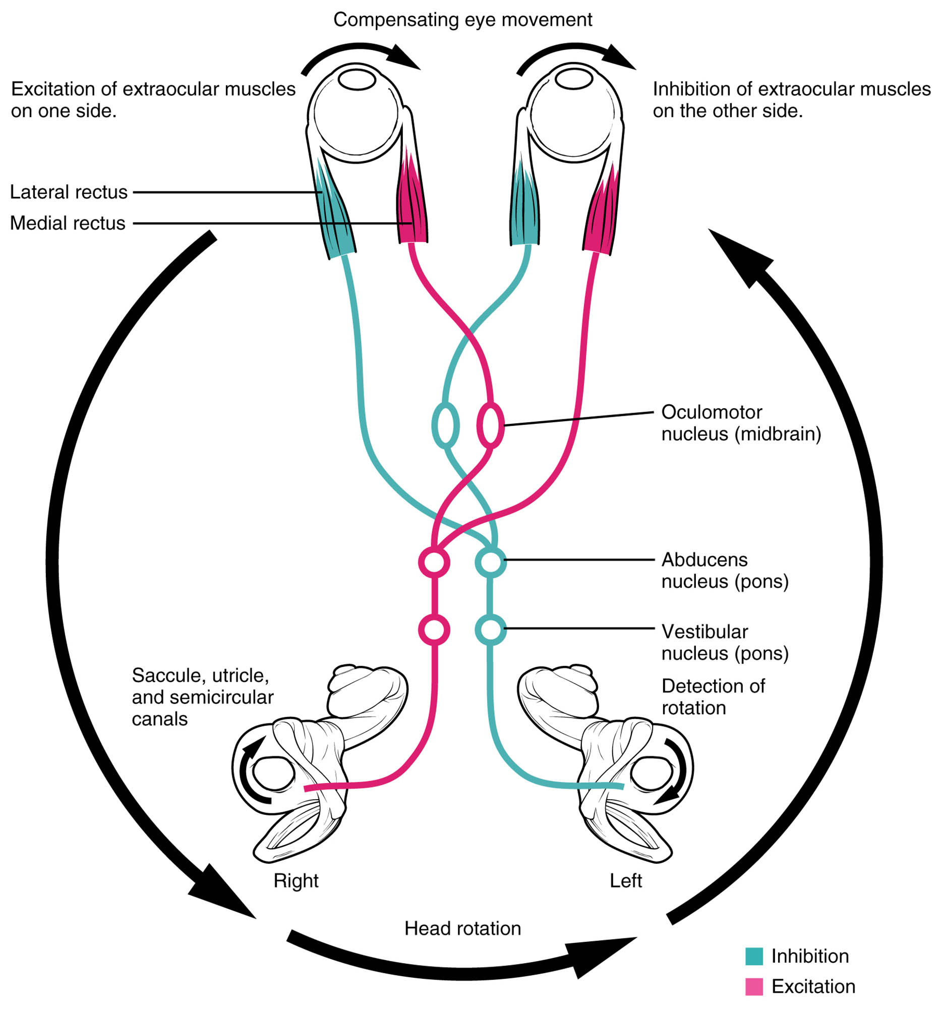
The vestibulo-ocular reflex (VOR) is a remarkable mechanism that maintains visual stability by coordinating the vestibular system with eye movements, even as the head moves. This diagram illustrates the neural connections and muscle actions that keep the eyes fixed on a target, countering head motion to ensure a steady field of view.
Vestibular nerve The vestibular nerve transmits signals from the vestibular system, including the semicircular canals and otolith organs, to the brainstem. It plays a critical role in detecting head movement and initiating the reflex to stabilize gaze.
Vestibular nuclei The vestibular nuclei, located in the medulla and pons, integrate vestibular input and coordinate with eye movement control centers. They relay signals to cranial nerve nuclei to adjust eye position during head motion.
Medial longitudinal fasciculus (MLF) The medial longitudinal fasciculus (MLF) is a fiber tract connecting the vestibular nuclei to the oculomotor, trochlear, and abducens nuclei. It ensures synchronized movement of both eyes, maintaining binocular vision during head turns.
Oculomotor nerve (III) The oculomotor nerve (III) innervates several extraocular muscles, including the medial rectus and superior rectus, to control eye movement. It receives signals from the vestibular nuclei to move the eyes opposite to head rotation.
Trochlear nerve (IV) The trochlear nerve (IV) supplies the superior oblique muscle, aiding in downward and outward eye movement. It works with the VOR to adjust eye position during head tilts and rotations.
Abducens nerve (VI) The abducens nerve (VI) controls the lateral rectus muscle, enabling outward eye movement. It collaborates with the VOR to counteract head movement and keep the visual target centered.
Medial rectus muscle The medial rectus muscle adducts the eye, moving it inward, and is activated during head turns to the opposite side. It helps maintain fixation on a target by opposing head rotation.
Lateral rectus muscle The lateral rectus muscle abducts the eye, moving it outward, and is engaged during head turns to the same side. It works to stabilize the gaze by counteracting lateral head motion.
Superior rectus muscle The superior rectus muscle elevates the eye and assists in inward rotation, activated during downward head movement. It ensures the visual field remains stable during vertical head adjustments.
Inferior rectus muscle The inferior rectus muscle depresses the eye and aids in outward rotation, responding to upward head movement. It contributes to keeping the target centered during upward head tilts.
Superior oblique muscle The superior oblique muscle rotates the eye downward and outward, activated during head tilts to the opposite side. It fine-tunes eye position to maintain visual fixation.
Inferior oblique muscle The inferior oblique muscle elevates and outwardly rotates the eye, engaged during head tilts to the same side. It supports the VOR by adjusting eye alignment during complex head movements.
Anatomy of the Vestibulo-Ocular Reflex
The vestibulo-ocular reflex involves a network of neural and muscular components that stabilize vision during head motion. This diagram outlines the anatomical connections that enable this reflex to function effectively.
- The vestibular nerve carries sensory input from the inner ear’s vestibular apparatus.
- The vestibular nuclei serve as the central hub, processing these signals.
- The medial longitudinal fasciculus (MLF) links these nuclei to cranial nerve motor centers.
- The oculomotor nerve (III), trochlear nerve (IV), and abducens nerve (VI) control the extraocular muscles.
- The medial rectus muscle, lateral rectus muscle, superior rectus muscle, inferior rectus muscle, superior oblique muscle, and inferior oblique muscle execute the compensatory movements.
- These structures work together to maintain a stable retinal image despite head movement.
Physiology of the Vestibulo-Ocular Reflex
The VOR operates by generating eye movements that oppose head motion, preserving visual focus. This diagram illustrates the physiological process behind this stabilizing reflex.
- The vestibular nerve detects angular or linear acceleration from head movement.
- The vestibular nuclei interpret these signals and send commands via the medial longitudinal fasciculus (MLF).
- The oculomotor nerve (III), trochlear nerve (IV), and abducens nerve (VI) activate the appropriate eye muscles.
- The medial rectus muscle and lateral rectus muscle adjust for horizontal head turns.
- The superior rectus muscle and inferior rectus muscle compensate for vertical movements.
- The superior oblique muscle and inferior oblique muscle handle torsional adjustments, ensuring precise gaze stability.
Role of Vestibular Input in Eye Stabilization
The vestibular nerve and vestibular nuclei are the sensory foundation of the VOR, detecting head motion. Their input drives the reflex to maintain visual fixation.
- The vestibular nerve relays signals from the semicircular canals and otolith organs.
- The vestibular nuclei integrate this data with eye position information.
- These nuclei adjust muscle activation based on head acceleration direction and speed.
- The reflex operates within milliseconds, ensuring real-time gaze stability.
- Damage to the vestibular nerve can disrupt balance and eye coordination.
- This system is particularly active during rapid head movements like walking or turning.
Role of Eye Muscles in Counteracting Motion
The extraocular muscles, controlled by cranial nerves, counteract head movement to stabilize vision. This diagram highlights their coordinated action in the VOR.
- The medial rectus muscle moves the eye inward during contralateral head turns.
- The lateral rectus muscle shifts the eye outward during ipsilateral head turns.
- The superior rectus muscle elevates the eye during downward head motion.
- The inferior rectus muscle depresses the eye during upward head motion.
- The superior oblique muscle and inferior oblique muscle adjust for torsional shifts.
- This muscular synergy ensures the retinal image remains steady, supporting clear vision.
Clinical Relevance of the Vestibulo-Ocular Reflex
Understanding the VOR’s anatomy and physiology aids in diagnosing and managing balance and eye movement disorders. This image provides a reference for assessing normal function.
- Damage to the vestibular nerve can lead to vestibular neuritis, causing vertigo and nystagmus.
- Vestibular nuclei dysfunction may result in impaired gaze stabilization and dizziness.
- Lesions in the medial longitudinal fasciculus (MLF) cause internuclear ophthalmoplegia.
- Weakness in the oculomotor nerve (III), trochlear nerve (IV), or abducens nerve (VI) can affect eye alignment.
- Muscle imbalances, such as in the medial rectus muscle, may lead to strabismus during head movement.
- VOR testing, like the head impulse test, evaluates reflex integrity.
- Rehabilitation exercises can improve VOR function in vestibular disorders.
In conclusion, the vestibulo-ocular reflex diagram reveals a sophisticated interplay between the vestibular system and eye muscles, ensuring visual stability during head movement. This elegant mechanism, driven by the vestibular nerve, vestibular nuclei, and coordinated muscle actions, highlights the body’s ability to adapt to dynamic environments, offering valuable insights into neurological and ocular health.

