The venous system plays an essential role in returning deoxygenated blood to the heart, relying on a unique anatomical design to manage low-pressure flow. This image presents a detailed sectional view of a vein, highlighting the tunica intima, tunica media, tunica adventitia, and valves, which together ensure efficient blood transport against gravity.
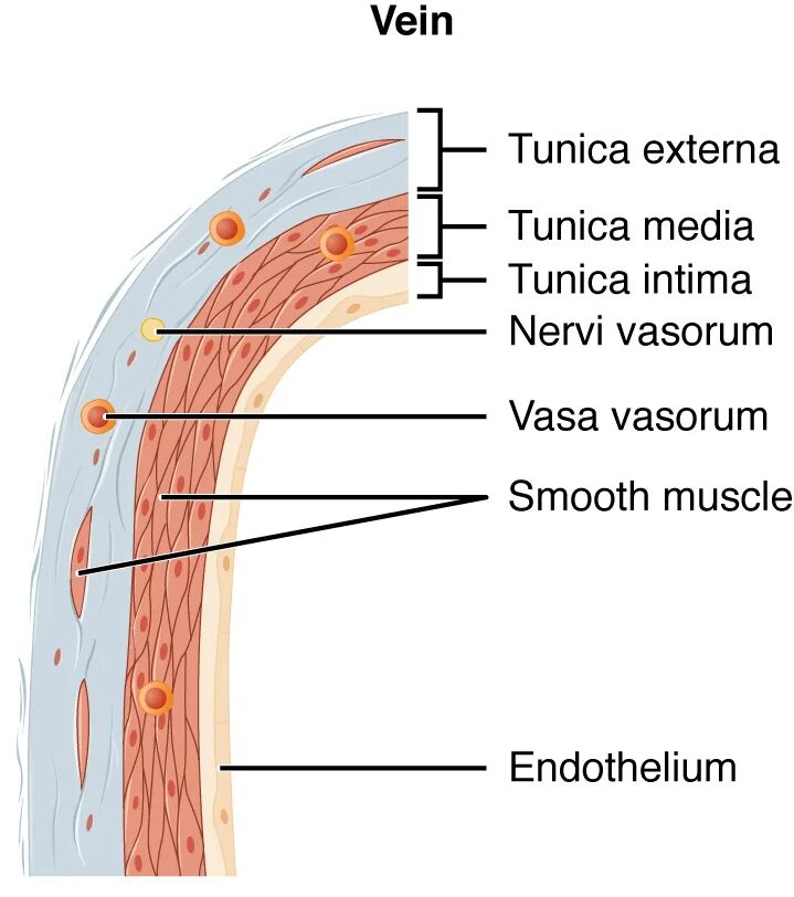
Tunica intima Tunica intima is the innermost layer of the vein, consisting of a thin endothelial lining that minimizes friction during blood flow. This layer includes a sparse subendothelial layer and lacks a prominent elastic lamina, reflecting the vein’s lower pressure environment.
Tunica media Tunica media forms the middle layer, comprising a thin layer of smooth muscle and elastic fibers that provide limited structural support. This thinner layer accommodates the low pressure of venous blood, typically around 10-15 mmHg, and relies on external compression for movement.
Tunica adventitia Tunica adventitia is the outermost and thickest layer, made up of dense connective tissue and collagen fibers that anchor the vein to surrounding tissues. This layer contains vasa vasorum and supports the vein’s structure, compensating for the weaker tunica media.
Valves Valves are flap-like structures within the vein that prevent backflow of blood, particularly in the lower extremities. These semilunar folds, composed of endothelial tissue, open with forward flow and close to maintain unidirectional movement toward the heart.
The Role of Venous Layers in Circulation
This examination highlights how each layer contributes to the vein’s function. The design supports the return of blood under low pressure and against gravity.
- Tunica Intima Function: The thin endothelial layer reduces resistance, facilitating smooth blood flow. Its flexibility allows veins to expand to accommodate larger blood volumes.
- Tunica Media Role: The minimal smooth muscle enables slight vasoconstriction but relies on skeletal muscle action for propulsion. This layer’s thinness reflects the vein’s passive role in circulation.
- Tunica Adventitia Support: The thick connective tissue provides structural stability, preventing collapse. The vasa vasorum ensures nutrient supply to the outer layers.
- Valve Mechanism: Valves ensure blood moves toward the heart, counteracting gravity. Their presence is critical in veins of the legs and arms.
Anatomical Details of Venous Structure
The sectional view reveals the vein’s layered construction, emphasizing its adaptations to low-pressure environments. The prominence of the tunica adventitia stands out in this design.
The image’s cross-sectional perspective offers a clear view of layer proportions. This understanding is vital for exploring venous health and functionality.
- Tunica Intima Composition: The endothelium is supported by a thin subendothelial layer, lacking the elastic lamina seen in arteries. This structure prioritizes flexibility over resilience.
- Tunica Media Thickness: This layer contains fewer smooth muscle cells and elastic fibers, making it thinner than in arteries. Its reduced thickness suits the vein’s lower pressure demands.
- Tunica Adventitia Variation: The outer layer is the thickest, rich in collagen to provide tensile strength. It also houses lymphatic vessels that drain excess interstitial fluid.
- Valve Anatomy: Valves are formed by folds of the tunica intima, reinforced by connective tissue. Their spacing increases in larger veins to manage greater blood volumes.
Physiological Functions of Venous Layers
The physiological roles of these layers are tailored to handle low-pressure blood return. Their design ensures efficient venous circulation throughout the body.
Each layer contributes to the vein’s ability to return blood. This functionality supports the body’s overall cardiovascular balance.
- Blood Volume Reservoir: Veins can expand to hold up to 60% of the body’s blood volume, aided by the thin tunica media. This capacity acts as a blood reservoir during hemorrhage.
- Venous Return: The tunica adventitia supports the vein’s structure during skeletal muscle contraction, the primary pump. Valves prevent reflux, ensuring steady flow to the heart.
- Oxygen and Waste Transport: Veins carry deoxygenated blood rich in carbon dioxide and metabolic waste. The tunica intima’s smoothness facilitates this transport.
- Pressure Regulation: The thin tunica media minimizes resistance, allowing low-pressure flow. Respiratory movements further assist in venous return.
Comparative Anatomy with Arteries
The venous structure contrasts with arteries, reflecting its distinct role in circulation. This sectional view emphasizes these differences clearly.
The image highlights the vein’s unique layering compared to arteries. This contrast is key to understanding circulatory dynamics.
- Venous vs. Arterial Walls: Veins have a thinner tunica media and thicker tunica adventitia, unlike the robust media of arteries. This reflects their lower pressure and reliance on external support.
- Valve Absence in Arteries: Valves are unique to veins, absent in arteries due to high, continuous pressure. This difference optimizes each vessel’s function.
- Elasticity Difference: Veins lack the extensive elastic lamellae of arteries, prioritizing compliance over recoil. This allows veins to accommodate variable blood volumes.
- Histological Contrast: Staining reveals less muscle in the tunica media and more collagen in the adventitia. This aids in microscopic differentiation from arteries.
Clinical and Research Perspectives
Insights from venous sectional views inform medical practice and research. The layered structure is a focus for studying venous health and pathology.
Advances in imaging and histology enhance these studies, offering diagnostic tools. These efforts support innovative treatment strategies.
- Varicose Veins Impact: Weak tunica media and valve incompetence lead to vein dilation. Sectional analysis assesses structural integrity for treatment planning.
- Deep Vein Thrombosis: Clots form in the tunica intima due to sluggish flow, a risk in immobile patients. Microscopic views guide anticoagulant therapy.
- Surgical Applications: Vein grafts require understanding layer thickness, especially the adventitia. This ensures compatibility in bypass procedures.
- Therapeutic Innovations: Strengthening the tunica media with compression therapy treats venous insufficiency. Stem cell research explores regenerating venous walls.
In conclusion, this image of a venous sectional view provides a detailed look at the tunica intima, tunica media, tunica adventitia, and valves, highlighting their critical roles in low-pressure blood return. These anatomical insights not only deepen our understanding of venous function but also support advancements in diagnosing and treating vascular conditions.

