Explore the complex network of veins that contribute to the superior vena cava with this insightful guide, featuring a detailed flow chart of venous drainage. This article delves into the anatomical structure, function, and clinical importance of these veins, offering a valuable resource for understanding upper body circulation.
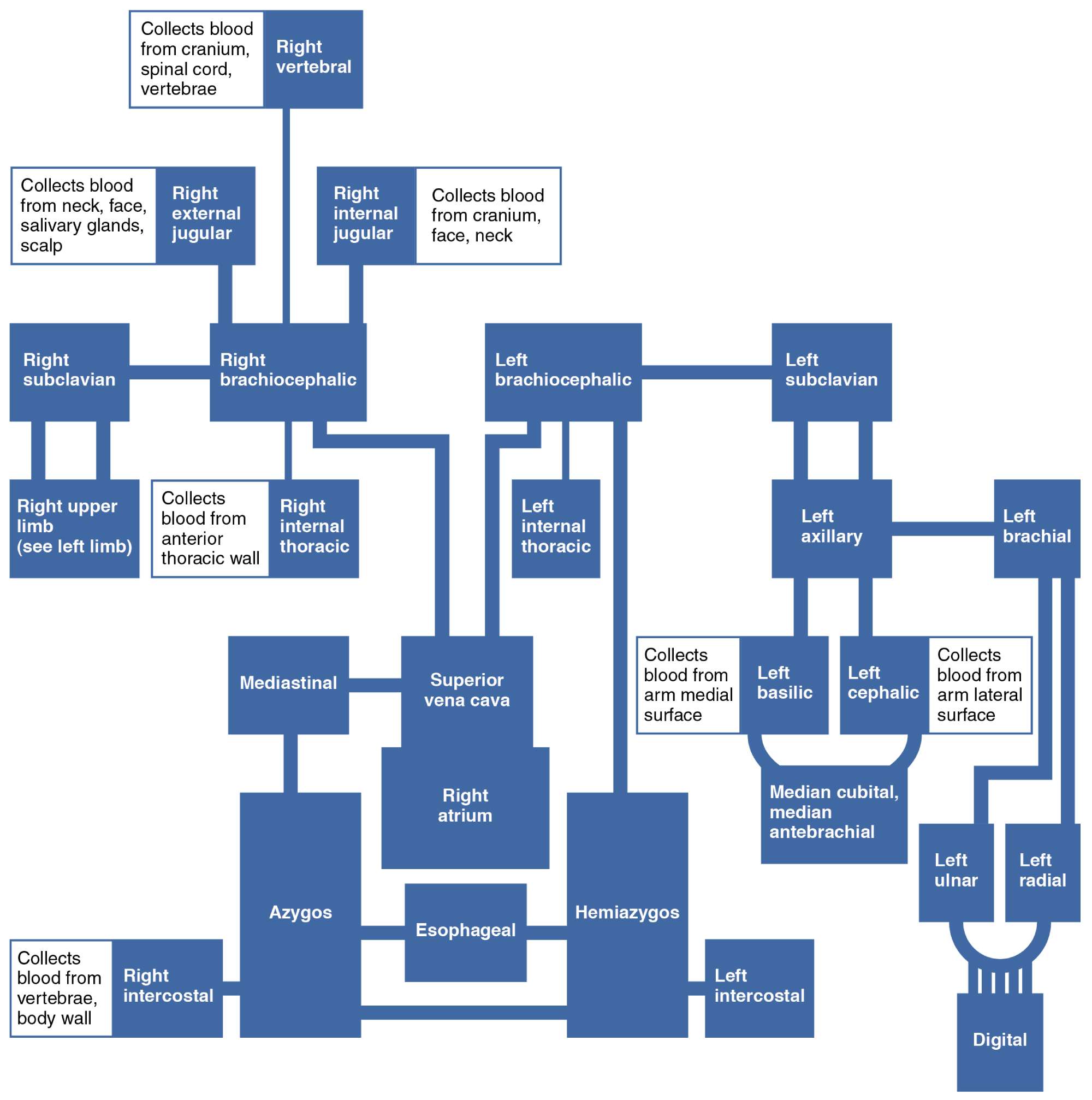
Right vertebral The right vertebral vein drains blood from the cranial region, including the spinal cord and vertebrae. It plays a key role in returning deoxygenated blood from the cervical spine to the systemic circulation.
Right external jugular The right external jugular vein collects blood from the face, neck, and scalp, including contributions from the salivary glands. It is a superficial vein that merges into the subclavian vein.
Right internal jugular The right internal jugular vein gathers blood from the cranium, face, and neck, serving as a major drainage route for the brain. It joins the subclavian vein to form the brachiocephalic vein.
Right brachiocephalic The right brachiocephalic vein is formed by the union of the internal jugular and subclavian veins, draining the right upper body. It transports blood into the superior vena cava.
Right subclavian The right subclavian vein receives blood from the upper limb via the axillary vein and contributes to the brachiocephalic vein. It is essential for draining the right arm and thoracic wall.
Right upper limb (see left limb) The right upper limb veins, similar to the left, collect blood from the arm and hand, feeding into the subclavian vein. This system mirrors the left side’s drainage pattern.
Right internal thoracic The right internal thoracic vein drains blood from the anterior thoracic wall, supporting the chest’s venous return. It empties into the brachiocephalic vein.
Mediastinal The mediastinal vein drains the mediastinum, the central compartment of the thoracic cavity. It contributes to the superior vena cava’s blood supply.
Superior vena cava The superior vena cava is a large vein that returns deoxygenated blood from the upper half of the body to the right atrium. It receives tributaries from the brachiocephalic veins and other thoracic veins.
Right atrium The right atrium receives blood from the superior vena cava, initiating the pulmonary circulation process. It is a critical chamber for processing deoxygenated blood.
Azygos The azygos vein drains the right intercostal spaces and thoracic wall, joining the superior vena cava. It provides an alternative drainage route in case of obstruction.
Esophageal The esophageal vein drains blood from the esophagus, contributing to the azygos system. It supports venous return from the digestive tract.
Hemiazygos The hemiazygos vein drains the left intercostal spaces, merging with the azygos vein. It complements the azygos system on the left side.
Left internal thoracic The left internal thoracic vein drains the left anterior thoracic wall, similar to its right counterpart. It feeds into the brachiocephalic vein.
Left brachiocephalic The left brachiocephalic vein forms from the left internal jugular and subclavian veins, draining the left upper body. It joins the right brachiocephalic to form the superior vena cava.
Left subclavian The left subclavian vein collects blood from the left upper limb via the axillary vein. It is a key contributor to the left brachiocephalic vein.
Left axillary The left axillary vein drains the left arm, receiving blood from the brachial and basilic veins. It continues as the subclavian vein.
Left brachial The left brachial vein collects blood from the medial arm surface, merging with the basilic vein. It is part of the deep venous system of the upper limb.
Left basilic The left basilic vein drains the medial arm and hand, joining the brachial vein. It is a common site for venous access.
Left cephalic The left cephalic vein collects blood from the lateral arm surface, feeding into the axillary vein. It is another accessible superficial vein.
Median cubital, median antebrachial The median cubital and median antebrachial veins connect the basilic and cephalic veins in the forearm. They are frequently used for phlebotomy.
Left ulnar The left ulnar vein drains the medial forearm, accompanying the ulnar artery. It merges with the radial vein to form the brachial vein.
Left radial The left radial vein drains the lateral forearm, following the radial artery. It contributes to the brachial vein system.
Digital The digital veins drain blood from the fingers, feeding into the palmar venous arches. They ensure efficient drainage from the digits.
Anatomical Overview of Veins into the Superior Vena Cava
The venous system flowing into the superior vena cava forms a critical pathway for upper body circulation. This network includes both deep and superficial veins that converge to return deoxygenated blood to the heart.
- The right vertebral and internal jugular veins handle cranial and facial drainage, essential for brain health.
- Major tributaries like the brachiocephalic veins integrate blood from both sides of the body.
- The azygos and hemiazygos veins provide additional drainage from the thoracic and intercostal regions.
- This system ensures efficient blood flow, supported by valves to prevent backflow.
Functional Roles and Clinical Relevance
The veins contributing to the superior vena cava serve vital physiological functions. Their clinical significance extends to various medical procedures.
- The subclavian and axillary veins are often used for central venous catheter placement.
- Obstructions in the superior vena cava can lead to superior vena cava syndrome, requiring prompt intervention.
- The digital and ulnar veins support hand circulation, critical in trauma cases.
- Understanding these pathways aids in diagnosing and treating circulatory disorders.
Physical Characteristics and Structure
The physical properties of these veins vary based on their location and function. This section explores their unique anatomy.
- The right atrium receives a high volume of blood, requiring robust venous input from the superior vena cava.
- Superficial veins like the cephalic and basilic are elastic and close to the skin, aiding accessibility.
- Deep veins such as the internal thoracic are paired with arteries, enhancing structural stability.
- Valves within the azygos and esophageal veins regulate flow against gravity.
Medical Applications and Considerations
Knowledge of these venous pathways is invaluable in medical practice. This section highlights their practical use.
- The median cubital vein is a preferred site for blood draws due to its size and location.
- Surgical planning often considers the left brachial and radial veins for vascular access.
- Monitoring the hemiazygos system helps manage thoracic trauma or obstructions.
- These insights support effective treatment of venous insufficiency or thrombosis.
Conclusion
The veins flowing into the superior vena cava create a sophisticated network essential for upper body venous return. From the digital veins in the fingers to the right atrium, each component plays a pivotal role in circulation. This guide offers a thorough understanding of their anatomy and clinical applications, encouraging further exploration to enhance medical expertise.

