The larynx, a pivotal structure in the human airway, extends from the laryngopharynx to the trachea, serving as both a protective gateway and the source of voice. Positioned below the hyoid bone, this cartilaginous organ facilitates respiration, phonation, and swallowing through its intricate design. An anterior view of the larynx offers a clear perspective on its components, enhancing understanding of its critical physiological roles.
Key Anatomical Labels in the Diagram
This section provides a detailed look at each labeled part, shedding light on their functions and positions within the larynx.
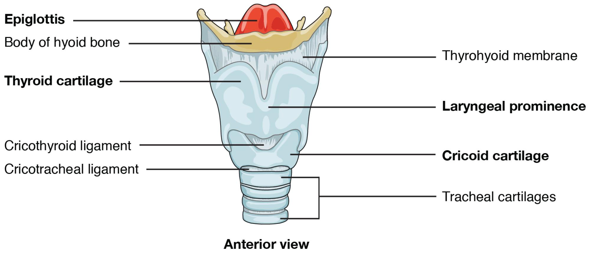
Epiglottis: The epiglottis is a flap of elastic cartilage that covers the laryngeal opening during swallowing, preventing food from entering the airway. It lifts during breathing to allow air to pass into the trachea.
Body of hyoid bone: The body of the hyoid bone is a horseshoe-shaped structure above the larynx, serving as an anchor for tongue and laryngeal muscles. It remains free-floating, supporting movements involved in speech and swallowing.
Thyroid cartilage: The thyroid cartilage forms the laryngeal framework, creating the prominent Adam’s apple that shields the vocal cords. It connects to the hyoid bone via the thyrohyoid membrane, providing structural support.
Cricothyroid ligament: The cricothyroid ligament links the thyroid cartilage to the cricoid cartilage, allowing tension adjustments for vocal pitch. It also serves as a potential site for emergency airway procedures.
Cricotracheal ligament: The cricotracheal ligament connects the cricoid cartilage to the trachea, ensuring a stable and flexible transition. It maintains airway integrity as air moves toward the lungs.
Thyrohyoid membrane: The thyrohyoid membrane stretches between the thyroid cartilage and hyoid bone, facilitating laryngeal elevation during swallowing. It houses the internal laryngeal nerve, which provides sensory input.
Laryngeal prominence: The laryngeal prominence, commonly known as the Adam’s apple, is the outward projection of the thyroid cartilage. It varies in size due to hormonal differences, offering protection to the vocal apparatus.
Cricoid cartilage: The cricoid cartilage is a ring-like structure below the thyroid cartilage, forming the larynx’s base. It provides a complete support ring, anchoring the airway and esophagus.
Tracheal cartilages: The tracheal cartilages are C-shaped rings extending from the cricoid cartilage, keeping the trachea open during respiration. They prevent collapse and guide air into the bronchial tree.
Cartilaginous and Ligamentous Structure
The larynx’s framework relies on a combination of cartilage and ligaments for stability and movement. This structure supports its dual roles effectively.
- The thyroid cartilage protects the vocal cords, with its prominence more pronounced in males.
- Cricoid cartilage offers a stable base, unique for its complete ring shape.
- The epiglottis acts as a dynamic flap, coordinating swallowing and breathing.
- Cricothyroid and cricotracheal ligaments ensure flexibility and continuity.
- Tracheal cartilages maintain airway patency under varying pressures.
Functional Roles in Respiration and Phonation
The larynx serves as a multifunctional hub, essential for breathing and voice production. Its anatomy enables a range of critical functions.
- The epiglottis prevents aspiration, directing food away from the trachea.
- Vocal cord vibration within the thyroid cartilage produces sound.
- The laryngeal prominence shields the delicate vocal apparatus.
- Tracheal cartilages ensure a clear airway to the lungs.
- The hyoid bone anchors muscles, supporting speech articulation.
Ligaments and Membranes in Action
Ligaments and membranes enhance the larynx’s mobility and stability. They connect key structures, allowing seamless operation.
- The thyrohyoid membrane elevates the larynx, aiding swallowing coordination.
- Cricothyroid ligament adjusts vocal pitch through muscle tension.
- Cricotracheal ligament maintains a smooth tracheal transition.
- These elements adapt to pressure changes, ensuring efficient airflow.
- The internal laryngeal nerve within the thyrohyoid membrane provides sensory feedback.
Clinical Relevance and Anatomical Considerations
Understanding laryngeal anatomy is crucial for diagnosing and treating related issues. Variations can guide clinical interventions.
- Laryngeal trauma may fracture the thyroid cartilage, affecting voice.
- Epiglottitis, an inflammation, can obstruct the airway, requiring urgent attention.
- Cricothyroid ligament access is used in emergency tracheostomies.
- Tracheal cartilage abnormalities may lead to breathing difficulties.
- Imaging studies help assess structural deviations for surgical planning.
The larynx’s elegant design, from the epiglottis to the tracheal cartilages, underscores its importance in respiration, phonation, and airway protection. By exploring its anatomy through the anterior view, one gains a deeper appreciation for its role in human physiology, highlighting the precision of this vital structure.

