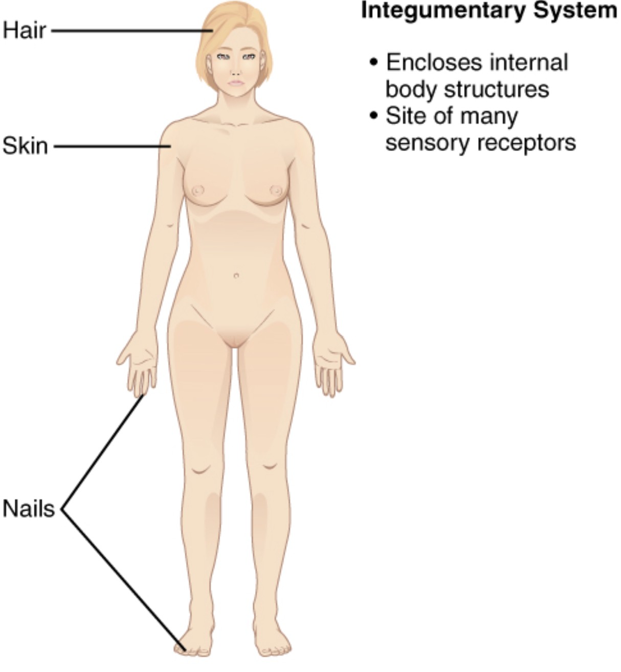
The human body’s largest organ system, the integumentary system, serves as a protective barrier and sensory interface with the environment. This image highlights the Epidermis, Dermis, Hypodermis, Hair Follicle, Sebaceous Gland, Sweat Gland, Arrector Pili Muscle, Sensory Nerve Ending, Blood Vessel, and Melanocyte, offering a comprehensive view of its layered structure and components. Exploring these elements reveals the system’s role in maintaining homeostasis and protecting internal organs.
Label Introductions:
- Epidermis: The epidermis is the outermost layer of the skin, acting as a waterproof barrier against pathogens and UV radiation. It continuously renews itself through cell division in its basal layer.
- Dermis: The dermis lies beneath the epidermis, providing strength and elasticity with collagen and elastin fibers. It houses blood vessels, nerves, and glands essential for skin function.
- Hypodermis: The hypodermis, or subcutaneous layer, connects the skin to underlying tissues, storing fat for insulation and energy. It also cushions organs and regulates temperature.
- Hair Follicle: The hair follicle is a tubular structure in the dermis where hair grows, anchoring it to the skin. It plays a role in temperature regulation and sensory perception.
- Sebaceous Gland: The sebaceous gland, attached to hair follicles, secretes sebum to keep skin and hair moisturized. It helps prevent dryness and protects against microbial growth.
- Sweat Gland: The sweat gland produces sweat to regulate body temperature through evaporation, located in the dermis or hypodermis. It also excretes small amounts of waste products.
- Arrector Pili Muscle: The arrector pili muscle is a small muscle that contracts to raise hair, causing goosebumps. It assists in trapping heat or signaling emotional responses.
- Sensory Nerve Ending: The sensory nerve ending detects touch, pressure, pain, and temperature, transmitting signals to the brain. It is distributed throughout the dermis for sensory feedback.
- Blood Vessel: The blood vessel in the dermis supplies nutrients and oxygen to skin cells while removing waste. It also aids in thermoregulation by adjusting blood flow.
- Melanocyte: The melanocyte, found in the epidermis, produces melanin to protect skin from UV damage. It determines skin pigmentation and varies in activity among individuals.
Introduction to the Integumentary System
The integumentary system encompasses the skin, hair, nails, and associated glands, forming a dynamic protective barrier. This system not only shields internal organs from external threats but also regulates temperature and facilitates sensory input. Its multilayered structure and specialized components work together to maintain overall health.
- Acts as the first line of defense against bacteria and injury.
- Regulates body temperature through sweat and blood flow.
- Synthesizes vitamin D when exposed to sunlight.
- Provides a medium for touch and pain sensation.
Epidermis: The Protective Outer Layer
The epidermis serves as the skin’s outermost defense, composed of stratified squamous epithelium. This layer prevents water loss and pathogen entry, with the stratum corneum acting as a tough barrier. Its continuous renewal ensures resilience against environmental wear.
- Contains keratinocytes that produce keratin for strength.
- Hosts melanocytes that offer UV protection via melanin.
- Undergoes desquamation to shed dead skin cells.
- Varies in thickness depending on body location.
Dermis: The Supportive Middle Layer
The dermis provides structural support beneath the epidermis, rich in connective tissue. It contains collagen for strength and elastin for flexibility, supporting skin’s resilience. This layer also houses critical structures like glands and nerves.
- Supplies nutrients via an extensive network of blood vessels.
- Contains mechanoreceptors for detecting pressure and vibration.
- Supports hair follicles and sebaceous glands.
- Thickens in areas subject to pressure, like the palms.
Hypodermis: The Deep Cushioning Layer
The hypodermis anchors the skin to underlying muscles and bones, serving as a fat storage site. This layer insulates the body, retaining heat, and cushions against physical impact. It also contains larger blood vessels and nerves for deeper tissue support.
- Varies in thickness based on gender and body region.
- Contributes to the body’s energy reserves during fasting.
- Facilitates lymphatic drainage and immune responses.
- Protects vital organs from external trauma.
Specialized Structures: Glands and Hair
The integumentary system features specialized structures like the hair follicle, sebaceous gland, and sweat gland. The hair follicle produces hair for protection and sensation, while the sebaceous gland secretes sebum for moisture. Sweat glands, including eccrine and apocrine types, regulate temperature and excrete waste.
- Sebaceous glands are more active during puberty due to hormonal changes.
- Sweat glands respond to heat or stress, releasing water and salts.
- Hair follicles cycle through growth, rest, and shedding phases.
- These structures maintain skin hydration and microbial balance.
Sensory and Vascular Components
The sensory nerve ending, blood vessel, and arrector pili muscle enhance the integumentary system’s functionality. Sensory nerve endings provide critical feedback on the environment, while blood vessels deliver oxygen and nutrients. The arrector pili muscle aids in thermoregulation and emotional signaling.
- Sensory endings include Pacinian corpuscles for deep pressure.
- Blood vessels dilate or constrict to adjust skin temperature.
- Arrector pili contraction traps air for warmth in cold conditions.
- These components integrate skin with the nervous system.
Melanocytes: Pigmentation and Protection
The melanocyte produces melanin, determining skin color and offering UV protection. Located in the basal layer of the epidermis, its activity increases with sun exposure to shield DNA. This process varies genetically, influencing diverse skin tones.
- Melanin absorbs UV rays to prevent skin cancer.
- Melanocyte activity is regulated by the hormone MSH.
- Overproduction can lead to hyperpigmentation disorders.
- Protects deeper skin layers from oxidative damage.
Conclusion
The integumentary system, with its epidermis, dermis, hypodermis, and specialized components, is a multifaceted organ system. From the protective role of melanocytes to the sensory capabilities of sensory nerve endings, it ensures the body’s resilience and adaptability. This understanding supports effective care and treatment of skin-related conditions, highlighting its importance in human health.

