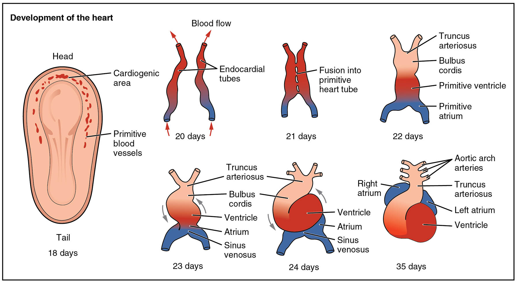
Discover the intricate process of how the human heart evolves from a simple structure at 18 days to a more defined organ by 35 days of embryonic development. This detailed exploration uses a comprehensive diagram to illustrate each critical stage, providing a window into the anatomical and physiological transformations that shape the cardiovascular system. From the initial formation of blood vessels to the emergence of distinct heart chambers, this guide offers a thorough understanding of this essential developmental journey.
Label Introduction
- Cardiogenic area The cardiogenic area is the initial site in the embryo where heart formation begins, located near the head end of the primitive streak. It develops into the region where the heart tube and early blood vessels originate.
- Primitive blood vessels Primitive blood vessels form the first network for blood circulation, emerging around the cardiogenic area by 18 days. These structures gradually evolve into the complex vascular system of the mature heart.
- Endocardial tubes Endocardial tubes are paired structures that appear at 20 days, which fuse to create the primitive heart tube. This fusion is a pivotal step in establishing the heart’s initial pumping mechanism.
- Fusion into primitive heart tube Fusion into primitive heart tube occurs at 21 days, merging the endocardial tubes into a single tubular structure. This tube begins to exhibit rhythmic contractions to support embryonic circulation.
- Truncus arteriosus The truncus arteriosus is the outflow tract of the heart tube, visible by 23 days, which will later divide into the aorta and pulmonary artery. It plays a crucial role in directing blood away from the heart.
- Bulbus cordis Bulbus cordis is a segment of the heart tube adjacent to the truncus arteriosus, noted at 23 days, contributing to the right ventricle and outflow tracts. Its proper development ensures effective blood ejection.
- Ventricle The ventricle forms as a dilated portion of the heart tube by 23 days, serving as the early pumping chamber. It will eventually split into the left and right ventricles during further development.
- Atrium The atrium emerges as a distinct region of the heart tube by 23 days, receiving blood from the sinus venosus. It will later divide into the left and right atria to handle dual circulation.
- Sinus venosus Sinus venosus is the inflow tract at 23 days, collecting blood from embryonic veins and delivering it to the atrium. It contributes to the sinoatrial node and parts of the right atrium in the mature heart.
- Right atrium The right atrium becomes recognizable by 35 days, receiving deoxygenated blood from the body via the vena cava. It is essential for the right side of the heart’s circulatory function.
- Ventricle This label, repeated at 24 days, reinforces the ventricle’s role as the pumping chamber, undergoing looping and differentiation. Its development is critical for the heart’s contractile ability.
- Atrium Noted again at 24 days, the atrium continues to develop as a receiving chamber, preparing for its division into two atria. This stage supports the heart’s growing complexity.
- Sinus venosus Reappearing at 24 days, the sinus venosus facilitates blood inflow, aiding the heart’s looping process. It remains vital for early circulation patterns.
- Left atrium The left atrium begins to form by 35 days, receiving oxygenated blood from the lungs via the pulmonary veins. Its development is key to separating oxygenated and deoxygenated blood.
- Aortic arch arteries Aortic arch arteries, visible by 35 days, are part of the arterial system that will remodel into the aorta and its branches. They support the transition to systemic circulation.
- Truncus arteriosus Seen again at 35 days, the truncus arteriosus continues to serve as the outflow tract, preparing for its division into major arteries. Its role is crucial for blood distribution.
Overview of Early Heart Development
The journey of heart development starts with the embryo at 18 days, marking the beginning of a complex process. This stage lays the foundation for the cardiovascular system, transforming simple tissues into a functional heart.
- Introduces the cardiogenic area as the starting point where heart formation initiates.
- Highlights the formation of primitive blood vessels to support early blood flow.
- Describes the initial appearance of endocardial tubes as precursors to the heart tube.
Formation of the Primitive Heart Tube
By 20 to 21 days, the heart begins to take shape with the fusion of key structures. This period is critical for establishing the heart’s basic pumping action.
- Explains the fusion of endocardial tubes into the primitive heart tube at 21 days.
- Details the onset of blood flow, driven by the tubular structure’s contractions.
- Notes the role of this early tube in supporting the embryo’s circulatory needs.
Looping and Differentiation of the Heart Tube
From 23 to 24 days, the heart tube undergoes looping, a vital step in its structural development. This process sets the stage for chamber formation.
- Describes the looping of the ventricle and atrium, guided by the sinus venosus.
- Highlights the emergence of the truncus arteriosus and bulbus cordis as outflow regions.
- Explains how this looping positions the heart for further specialization.
Emergence of Atrial and Ventricular Regions
By 35 days, the heart starts to define its atrial and ventricular regions. This differentiation is essential for the heart’s future four-chambered structure.
- Details the development of the right atrium and left atrium, receiving different blood types.
- Discusses the ventricle‘s role as the primary pumping chamber, preparing for division.
- Notes the contribution of the aortic arch arteries to the emerging vascular network.
Role of Outflow Tracts in Heart Development
The outflow tracts, including the truncus arteriosus, are critical for directing blood flow. Their development ensures the heart can handle increasing circulatory demands.
- Explains how the truncus arteriosus will split into the aorta and pulmonary artery.
- Describes the bulbus cordis contribution to the right ventricle and outflow tracts.
- Highlights the remodeling of aortic arch arteries into major arterial branches.
Physiological Implications of Early Heart Stages
The early stages of heart development have lasting effects on its function. Understanding these phases provides insight into congenital heart conditions.
- Notes how the sinus venosus evolves into the sinoatrial node, regulating heart rhythm.
- Discusses the atrium and ventricle differentiation as a foundation for chamber separation.
- Emphasizes the importance of proper truncus arteriosus division to avoid defects.
Clinical Considerations in Heart Embryology
Knowledge of these developmental stages aids in recognizing potential abnormalities. Early interventions can address issues arising from improper formation.
- Identifies risks of truncus arteriosus malformation leading to a single outflow vessel.
- Explains how bulbus cordis anomalies can affect ventricular development.
- Highlights the significance of aortic arch arteries remodeling for normal circulation.
In conclusion, the embryological development of the human heart from 18 to 35 days is a remarkable transformation from a simple tube to a structured organ. Each stage, from the cardiogenic area to the defined right atrium and left atrium, builds the foundation for a robust cardiovascular system. This diagram serves as an invaluable resource for understanding the anatomical and physiological evolution of the heart, offering insights into both normal development and potential congenital variations.

