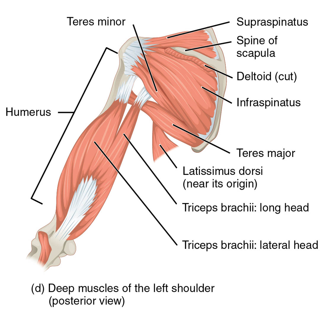
The shoulder’s posterior region is a critical area of the human body, housing a sophisticated array of muscles that drive movement and maintain stability. This article examines the deep muscles of the left shoulder as depicted in the provided medical image, focusing on the humerus, teres minor, supraspinatus, spine of scapula, deltoid, infraspinatus, teres major, latissimus dorsi, triceps brachii: long head, and triceps brachii: lateral head. These structures are essential for understanding upper body mechanics and their role in daily activities. By exploring their anatomy and functions, readers can gain a deeper appreciation of the shoulder’s complex design and its importance in physical health.
Introduction to the Labeled Muscles
This section provides a clear overview of the muscles and structures labeled in the image, laying a strong foundation. The following details break down each component to enhance your grasp of posterior shoulder anatomy.
- Humerus: This long bone of the upper arm serves as the primary attachment point for shoulder muscles. It facilitates arm movement and supports the shoulder joint’s structural integrity.
- Teres minor: Located near the scapula, this small muscle aids in external rotation of the arm. It is a key component of the rotator cuff, stabilizing the shoulder during motion.
- Supraspinatus: Positioned above the spine of the scapula, this muscle initiates arm abduction. It plays a vital role in the rotator cuff, protecting the shoulder joint.
- Spine of scapula: This bony ridge on the scapula provides attachment sites for muscles. It enhances the scapula’s stability and supports upper back structure.
- Deltoid: Shown as a cut muscle over the shoulder, it covers the joint and enables arm abduction. Its posterior fibers contribute to extension and rotation.
- Infraspinatus: Found below the spine of the scapula, this muscle externally rotates the arm. It is another essential rotator cuff muscle for shoulder stability.
- Teres major: Positioned below the shoulder, this muscle assists in arm adduction and extension. It works with the latissimus dorsi for powerful pulling actions.
- Latissimus dorsi: Visible near its origin, this broad muscle extends and adducts the arm. It originates from the thoracolumbar fascia, aiding in back and arm strength.
- Triceps brachii: long head: This head of the triceps extends the elbow and stabilizes the shoulder. It originates from the infraglenoid tubercle of the scapula.
- Triceps brachii: lateral head: Located on the outer arm, this head extends the elbow. It works with the long head to enhance arm extension and strength.
Detailed Anatomical Overview
This part dives into the structural details of these muscles, offering a thorough examination. The humerus, teres minor, supraspinatus, spine of scapula, deltoid, infraspinatus, teres major, latissimus dorsi, triceps brachii: long head, and triceps brachii: lateral head form a cohesive unit in the posterior shoulder.
- The humerus articulates with the scapula at the glenohumeral joint, receiving muscle insertions like the deltoid tuberosity. Its robust structure supports weight-bearing and movement.
- The teres minor originates from the lateral border of the scapula, inserting into the greater tubercle of the humerus. Innervated by the axillary nerve, it ensures smooth rotation.
- The supraspinatus arises from the supraspinous fossa, inserting into the humerus’ greater tubercle. Its innervation by the suprascapular nerve is critical for initial abduction.
- The spine of scapula separates the supraspinatus and infraspinatus fossae, providing leverage. It serves as an attachment for the trapezius and deltoid muscles.
- The deltoid originates from the spine of the scapula and clavicle, inserting into the deltoid tuberosity. Its axillary nerve supply supports its multifaceted role.
- The infraspinatus originates from the infraspinous fossa, inserting into the humerus. Innervated by the suprascapular nerve, it enhances external rotation and stability.
- The teres major originates from the inferior angle of the scapula, inserting into the humerus’ intertubercular groove. Its lower subscapular nerve innervation aids extension.
- The latissimus dorsi extends from the thoracolumbar fascia to the humerus, innervated by the thoracodorsal nerve. Its broad surface enhances pulling strength.
- The triceps brachii: long head originates from the scapula’s infraglenoid tubercle, inserting into the olecranon. Its radial nerve supply supports elbow extension.
- The triceps brachii: lateral head arises from the posterior humerus, also inserting into the olecranon. It complements the long head for powerful arm extension.
Functional Roles in the Body
This section explores how these muscles contribute to movement and stability, providing practical insights. The humerus, teres minor, supraspinatus, spine of scapula, deltoid, infraspinatus, teres major, latissimus dorsi, triceps brachii: long head, and triceps brachii: lateral head are integral to posterior shoulder function.
- The humerus acts as a lever for muscle action, enabling lifting and pulling. Its alignment with the scapula ensures efficient force transmission.
- The teres minor stabilizes the shoulder during overhead activities like throwing. Its rotator cuff role prevents joint dislocation under stress.
- The supraspinatus initiates the first 15 degrees of arm abduction, crucial for reaching. It works with the deltoid for full range of motion.
- The spine of scapula provides a structural base for muscle attachment, enhancing scapular movement. It supports posture during upper body exercises.
- The deltoid powers arm lifting and extension, with posterior fibers active in pulling. Its strength is vital for athletic performance.
- The infraspinatus ensures smooth external rotation, essential for sports like tennis. It maintains joint integrity during repetitive motions.
- The teres major supports rowing and climbing by aiding arm extension. Its synergy with the latissimus dorsi boosts upper body power.
- The latissimus dorsi drives swimming and pulling motions, enhancing endurance. Its extensive origin provides a wide range of motion.
- The triceps brachii: long head extends the elbow during pushing, stabilizing the shoulder. It prevents hyperextension during heavy lifts.
- The triceps brachii: lateral head strengthens elbow extension, aiding in pressing. It complements the long head for balanced arm function.
Clinical Significance and Considerations
This part addresses the medical relevance of these muscles, offering insights into potential issues. The humerus, teres minor, supraspinatus, spine of scapula, deltoid, infraspinatus, teres major, latissimus dorsi, triceps brachii: long head, and triceps brachii: lateral head are susceptible to specific conditions.
- The humerus can fracture from trauma, affecting shoulder function. Proper immobilization and physical therapy aid recovery.
- The teres minor may tear in rotator cuff injuries, causing pain. Rehabilitation focuses on restoring rotation and stability.
- The supraspinatus is prone to tendinitis from overuse, limiting abduction. Rest and anti-inflammatory treatments are common solutions.
- The spine of scapula can be involved in scapular fractures, impacting mobility. Surgical intervention may be required in severe cases.
- The deltoid may suffer strains from overexertion, reducing strength. Targeted exercises help regain full function.
- The infraspinatus can develop tears, leading to weakness. Physical therapy strengthens the rotator cuff to prevent recurrence.
- The teres major may experience overuse injuries, causing discomfort. Stretching and rest promote healing.
- The latissimus dorsi can strain from heavy lifting, affecting back motion. Gradual strengthening aids recovery.
- The triceps brachii: long head may rupture, impairing elbow extension. Surgical repair may be necessary for severe cases.
- The triceps brachii: lateral head can develop tendinitis, causing pain. Rest and therapy alleviate symptoms effectively.
As we conclude this journey, the humerus, teres minor, supraspinatus, spine of scapula, deltoid, infraspinatus, teres major, latissimus dorsi, triceps brachii: long head, and triceps brachii: lateral head reveal the intricate beauty of the posterior shoulder. Their coordinated efforts enable a wide range of motions, from lifting to throwing, while maintaining joint stability. This knowledge not only enriches anatomical understanding but also guides effective management of shoulder-related conditions, encouraging further exploration of their nerve and vascular networks.

