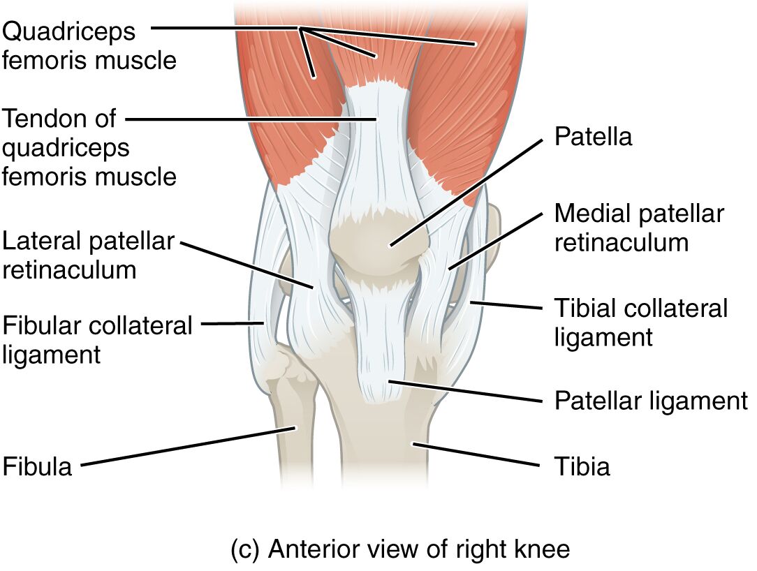
The knee joint, the largest hinge joint in the human body, serves as a cornerstone for mobility and weight support in daily activities. This anterior view of the right knee highlights the intricate network of muscles, ligaments, and bones that work together to ensure stability and movement. Understanding this anatomical layout provides valuable insights into the knee’s function and the importance of its structural components.
Labels Introduction
- Quadriceps femoris muscle: The quadriceps femoris muscle, located on the front of the thigh, is a powerful extensor of the knee joint, crucial for activities like standing and walking. It consists of four heads that converge into a common tendon, providing significant strength and stability.
- Tendon of quadriceps femoris muscle: This tendon connects the quadriceps muscle to the patella, transmitting force to extend the knee during movement. It plays a vital role in maintaining upright posture and is susceptible to strain with overuse.
- Patella: The patella, or kneecap, is a sesamoid bone that protects the knee joint and increases the leverage of the quadriceps muscle. It glides within a femoral groove, facilitating smooth knee flexion and extension.
- Medial patellar retinaculum: The medial patellar retinaculum provides lateral support to the patella, stabilizing it during knee motion. It consists of fibrous tissue that connects the patella to surrounding structures, reducing the risk of dislocation.
- Lateral patellar retinaculum: Located on the outer side, the lateral patellar retinaculum supports the patella’s alignment and prevents excessive lateral movement. It works in tandem with the medial retinaculum to maintain patellar tracking.
- Fibular collateral ligament: The fibular collateral ligament runs along the lateral side of the knee, providing stability against varus forces that could bend the joint inward. It connects the femur to the fibula, enhancing lateral support.
- Tibial collateral ligament: The tibial collateral ligament, on the medial side, resists valgus forces and stabilizes the knee against inward bending. It is broader and stronger than the fibular ligament, anchoring the femur to the tibia.
- Patellar ligament: The patellar ligament extends from the patella to the tibia, transmitting quadriceps force to straighten the knee. It is a continuation of the quadriceps tendon and is critical for jumping and running.
- Fibula: The fibula, a slender bone parallel to the tibia, provides lateral support to the knee and ankle joints. It serves as an attachment point for ligaments and muscles, contributing to overall leg stability.
- Tibia: The tibia, or shin bone, forms the weight-bearing lower part of the knee joint, articulating with the femur. It supports the body’s mass and provides a stable base for the knee’s hinge mechanism.
Anatomical and Physical Introduction
The anterior view of the right knee showcases a remarkable assembly of structures that enable its dual role as a weight-bearing and mobile joint. The quadriceps femoris muscle, with its robust tendon, drives knee extension, essential for standing and locomotion. The patella enhances this action by acting as a fulcrum, amplifying the muscle’s power while shielding the joint.
Ligaments such as the fibular and tibial collateral ligaments fortify the knee’s lateral stability, resisting forces that could misalign the joint. The patellar ligament, a key extension component, ensures efficient force transmission from the quadriceps to the tibia, vital for dynamic movements. Retinacula on both medial and lateral sides reinforce patellar alignment, preventing tracking issues that could lead to pain or injury.
The fibula and tibia together form a sturdy framework, with the tibia bearing most of the load and the fibula offering supplementary support. This structural synergy allows the knee to adapt to various stresses, from walking to athletic endeavors. The absence of disease in this image focuses attention on the healthy anatomy, emphasizing the importance of maintaining these components through proper care and exercise.
Conclusion
The anterior view of the right knee joint reveals the elegant interplay of muscles, ligaments, and bones that sustain its function. Each element, from the quadriceps to the tibial collateral ligament, contributes to a resilient system capable of withstanding daily demands. Appreciating this anatomy fosters a deeper understanding of knee health and the need to protect it from potential wear or trauma.

