Antigen processing and presentation are essential steps in the immune system, enabling the body to detect and respond to foreign invaders with precision. This process involves breaking down antigens into smaller peptides and displaying them on the surface of cells via major histocompatibility complex (MHC) molecules, where they are recognized by T cells. This detailed illustration provides a clear view of the mechanisms involved, shedding light on how the immune system coordinates its adaptive responses.
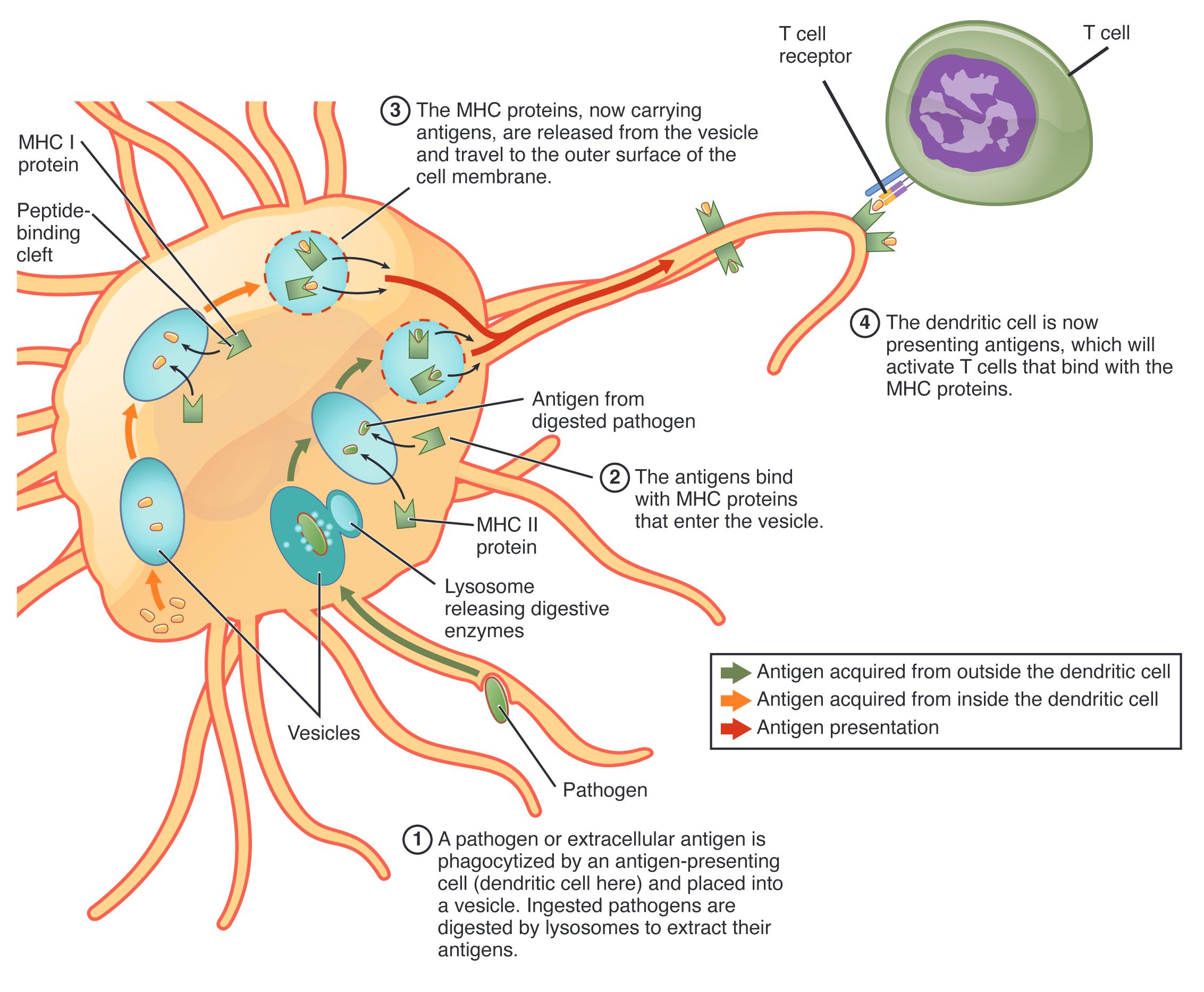
Labeled Components of Antigen Processing and Presentation
Antigen: This foreign substance, such as a protein from a pathogen, is taken up by antigen-presenting cells for processing. It is broken down into peptides that are presented to T cells to trigger an immune response.
Endocytosis: This process allows antigen-presenting cells to engulf antigens from the extracellular environment. It initiates the degradation of antigens into smaller fragments.
Lysosome: Containing digestive enzymes, the lysosome fuses with the endocytic vesicle to break down antigens. This step generates peptide fragments for presentation.
Endocytic vesicle: Formed during endocytosis, this vesicle transports antigens into the cell for processing. It merges with lysosomes to facilitate antigen degradation.
Peptide fragment: These small antigen pieces, produced by lysosomal enzymes, are loaded onto MHC molecules. They serve as the specific targets recognized by T cell receptors.
MHC class II molecule: Found on antigen-presenting cells like macrophages and dendritic cells, this molecule binds peptide fragments. It presents them on the cell surface for recognition by CD4+ T cells.
Endoplasmic reticulum (ER): This organelle is involved in the assembly of MHC class I molecules within the cell. It provides a site for loading peptides derived from intracellular antigens.
Proteasome: This cellular structure degrades intracellular proteins, including viral or damaged proteins, into peptides. It supplies peptide fragments for MHC class I presentation.
MHC class I molecule: Present on nearly all nucleated cells, this molecule binds peptides from intracellular sources. It displays them on the cell surface for recognition by CD8+ T cells.
Golgi apparatus: This organelle modifies and packages MHC-peptide complexes for transport to the cell surface. It ensures proper presentation of antigens to T cells.
Cell membrane: The outer layer of the cell where MHC-peptide complexes are displayed for T cell recognition. It serves as the interface between the cell and the immune system.
CD4+ T cell: This helper T cell recognizes MHC class II-peptide complexes, activating other immune cells. It coordinates the adaptive immune response against extracellular pathogens.
CD8+ T cell: This cytotoxic T cell binds to MHC class I-peptide complexes, targeting infected or abnormal cells. It directly kills cells harboring intracellular pathogens.
Antigen-presenting cell (APC): Cells like dendritic cells, macrophages, and B cells process and present antigens via MHC molecules. They are crucial for initiating T cell responses.
Anatomical Context of Antigen Processing
Antigen processing occurs within specialized cellular compartments, ensuring effective immune activation.
- Antigens are internalized via endocytosis, entering endocytic vesicles.
- Lysosomes break down antigens, producing peptide fragments for presentation.
- The endoplasmic reticulum assembles MHC class I molecules with intracellular peptides.
- Proteasomes degrade cytosolic proteins, supplying peptides for MHC class I.
- The Golgi apparatus refines MHC-peptide complexes for surface display.
- The cell membrane presents these complexes to circulating T cells.
This illustration highlights the intracellular journey of antigens.
Physiological Role in Immune Response
Antigen processing and presentation bridge innate and adaptive immunity effectively.
- Endocytosis captures extracellular antigens, initiating the MHC class II pathway.
- Lysosomal enzymes generate peptide fragments, essential for MHC loading.
- MHC class II molecules present peptides to CD4+ T cells, boosting helper functions.
- The proteasome supplies peptides from intracellular sources for MHC class I.
- MHC class I complexes activate CD8+ T cells, targeting infected cells.
- Antigen-presenting cells amplify the response by engaging multiple T cells.
This process ensures a tailored immune attack on pathogens.
Mechanisms of MHC Presentation
The MHC system employs distinct pathways for different antigen sources.
- MHC class II molecules bind peptides from endocytic vesicles, targeting extracellular threats.
- The endoplasmic reticulum loads MHC class I with peptides from the proteasome.
- The Golgi apparatus ensures proper trafficking of MHC complexes to the membrane.
- CD4+ T cells recognize MHC class II, enhancing antibody production.
- CD8+ T cells bind MHC class I, triggering apoptosis in infected cells.
- Antigen-presenting cells optimize peptide presentation for T cell activation.
This dual pathway reflects the immune system’s versatility.
Clinical Relevance of Antigen Processing
Understanding this process aids in managing immune-related disorders.
- Defects in MHC class II presentation can lead to immunodeficiency, reducing CD4+ T cell activity.
- MHC class I dysfunction may impair CD8+ T cell responses, increasing viral persistence.
- Endocytosis or lysosomal issues can hinder antigen processing, weakening immunity.
- Proteasome abnormalities are linked to cancer, where cells evade T cell detection.
- Antigen-presenting cell dysfunction is studied in autoimmune diseases.
- MHC typing guides organ transplant compatibility by assessing antigen presentation.
This knowledge supports targeted therapeutic strategies.
Developmental and Adaptive Features
Antigen processing adapts to the body’s changing needs over time.
- Antigen-presenting cells mature, enhancing endocytosis and presentation efficiency.
- Lysosomes and proteasomes refine their activity with immune experience.
- MHC molecules develop diversity through genetic variation, broadening antigen coverage.
- The endoplasmic reticulum and Golgi adapt to intracellular pathogen challenges.
- CD4+ and CD8+ T cells gain specificity through thymic selection.
- This adaptability ensures robust responses to evolving pathogens.
This evolution strengthens long-term immune protection.
Antigen processing and presentation, as illustrated, are vital for linking antigen recognition to immune activation, with MHC molecules and T cells working in unison. By breaking down antigens and displaying them on cell surfaces, this process enables the body to mount a precise and effective defense, making it a captivating area of study for those interested in immunology and health.

