The inferior vena cava plays a crucial role in the circulatory system by collecting deoxygenated blood from the lower body and returning it to the heart. This comprehensive guide explores the intricate network of veins that contribute to this process, highlighting key anatomical structures and their physiological significance for efficient blood flow.
Key Labels in the Venous Flowchart
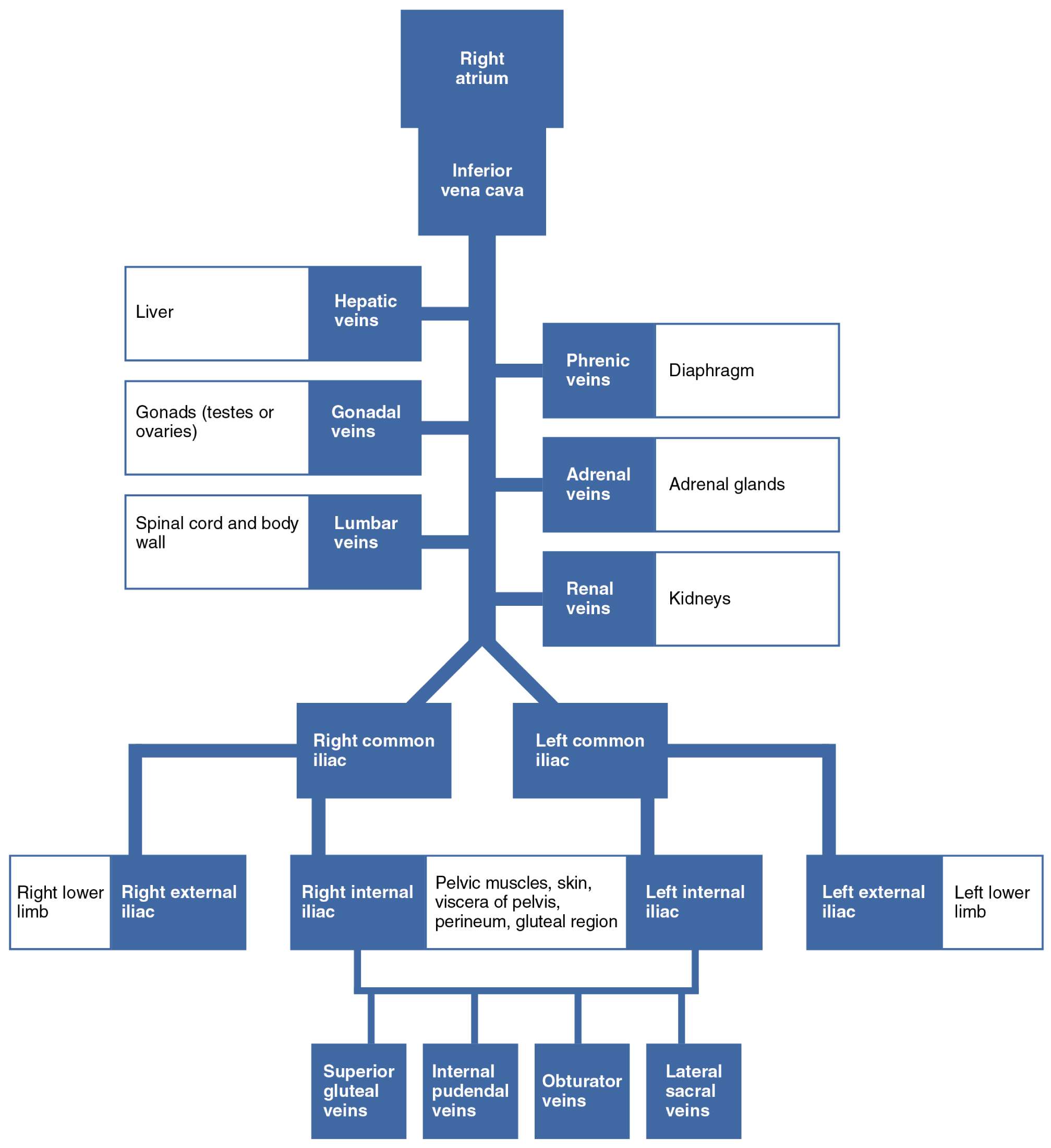
The flowchart illustrates the hierarchical organization of veins draining into the inferior vena cava. Each label represents a specific anatomical component involved in venous return.
Right atrium The right atrium is the upper chamber of the heart that receives deoxygenated blood from the body. It serves as the entry point for blood returning via the inferior vena cava before being pumped to the lungs for oxygenation.
Inferior vena cava The inferior vena cava is the largest vein in the body, responsible for transporting deoxygenated blood from the lower extremities, pelvis, and abdomen to the heart. It forms from the union of the common iliac veins and ascends through the abdomen to empty into the right atrium.
Liver The liver is a vital organ involved in metabolism, detoxification, and nutrient processing. It receives nutrient-rich blood from the portal system, and its deoxygenated blood drains back to the inferior vena cava through specific veins.
Hepatic veins Hepatic veins collect blood from the liver’s sinusoids after processing. These veins ensure that metabolized substances are returned to systemic circulation, playing a key role in maintaining homeostasis.
Phrenic veins Phrenic veins drain blood from the diaphragm, the primary muscle of respiration. They contribute to the inferior vena cava, supporting the venous return from structures involved in breathing mechanics.
Diaphragm The diaphragm is a dome-shaped muscle separating the thoracic and abdominal cavities, essential for inhalation. Its venous drainage helps prevent congestion in the respiratory apparatus during physical activity.
Gonads (testes or ovaries) The gonads are reproductive organs that produce gametes and hormones such as testosterone or estrogen. Their venous drainage is asymmetric, with the right gonadal vein entering the inferior vena cava directly and the left typically joining the renal vein.
Gonadal veins Gonadal veins transport deoxygenated blood from the gonads back to the central circulation. Variations in their anatomy can influence conditions like varicocele in males or pelvic congestion in females.
Spinal cord and body wall The spinal cord and body wall encompass neural tissues and muscular structures along the vertebral column. Their venous network supports nutrient delivery and waste removal from these critical areas.
Lumbar veins Lumbar veins drain the lower back, including muscles and vertebrae. They form connections with the azygos system, providing alternative pathways for venous return in case of obstructions.
Adrenal veins Adrenal veins collect blood from the adrenal glands after hormone secretion. The right adrenal vein drains directly into the inferior vena cava, while the left often joins the renal vein.
Adrenal glands Adrenal glands are endocrine organs atop the kidneys that release hormones like cortisol and aldosterone for stress response and electrolyte balance. Their venous outflow ensures rapid distribution of these hormones systemically.
Renal veins Renal veins carry filtered blood from the kidneys back to the inferior vena cava. They are essential for removing waste products like urea and maintaining fluid balance.
Kidneys The kidneys filter blood to produce urine, regulating electrolytes and blood pressure. Each kidney’s venous system handles a significant portion of cardiac output for efficient filtration.
Right common iliac The right common iliac vein forms from the union of external and internal iliac veins on the right side. It transports blood from the right lower limb and pelvis to the inferior vena cava.
Left common iliac The left common iliac vein mirrors its right counterpart, collecting from the left pelvis and lower limb. It joins the right to form the inferior vena cava at the L5 vertebral level.
Right lower limb The right lower limb includes structures from the hip to the foot, involved in locomotion. Its venous return prevents pooling and supports overall circulation during movement.
Right external iliac The right external iliac vein drains the lower limb, continuing from the femoral vein. It handles blood from superficial and deep tissues, crucial for preventing edema.
Right internal iliac The right internal iliac vein collects from pelvic organs and muscles. It branches into multiple tributaries to accommodate the complex pelvic anatomy.
Pelvic muscles, skin, viscera of pelvis, perineum, gluteal region This region encompasses supportive muscles, integument, and organs like the bladder and rectum. Venous drainage here prevents congestion that could affect reproductive and excretory functions.
Left internal iliac The left internal iliac vein mirrors the right, draining pelvic structures. It supports blood return from organs involved in digestion and reproduction.
Left external iliac The left external iliac vein collects from the left lower limb. It ensures efficient deoxygenated blood transport to maintain tissue health.
Left lower limb The left lower limb parallels the right in function and venous needs. Proper drainage is vital for mobility and preventing conditions like deep vein thrombosis.
Superior gluteal veins Superior gluteal veins drain the gluteus maximus and surrounding muscles. They contribute to pelvic venous return, aiding in posture and gait.
Internal pudendal veins Internal pudendal veins serve the perineum and external genitalia. They are important for sensory and erectile tissues in both sexes.
Obturator veins Obturator veins drain the thigh adductor muscles and hip joint. They connect to the internal iliac system, supporting lower body movement.
Lateral sacral veins Lateral sacral veins collect from the sacrum and coccyx regions. They form part of the sacral venous plexus, linking to spinal veins.
The Role of the Inferior Vena Cava in Systemic Circulation
The inferior vena cava acts as a major conduit for venous blood from below the diaphragm. Understanding its tributaries enhances comprehension of circulatory dynamics and potential clinical issues.
- It originates at the L5 vertebra from the confluence of the common iliac veins.
- The vessel ascends retroperitoneally, piercing the diaphragm at T8 to enter the right atrium.
- Physiologically, it handles about 70% of venous return, influenced by abdominal pressure and respiration.
- Valves are minimal, relying on muscle pumps for flow against gravity.
Major Tributaries and Their Contributions
Tributaries to the inferior vena cava are categorized as paired or unpaired based on symmetry. This organization reflects embryonic development and functional efficiency.
- Hepatic veins typically number three (right, middle, left), draining liver lobes after portal processing.
- Renal veins lie anterior to the arteries, with the left longer and crossing the aorta.
- Gonadal veins show sexual dimorphism in drainage patterns, impacting varicose vein risks.
- Lumbar veins, usually four pairs, communicate with vertebral plexuses for collateral circulation.
- Adrenal veins ensure hormone-laden blood reaches the heart quickly.
- Phrenic veins accompany the phrenic arteries, draining the diaphragmatic undersurface.
Physiological Importance of Venous Drainage
Efficient venous drainage prevents stasis and supports organ function. Disruptions can lead to edema or thrombosis, underscoring the system’s resilience.
- In the lower limbs, external iliac veins rely on calf muscle contractions as a “peripheral heart.”
- Internal iliac branches, like superior gluteal veins, facilitate blood return during sitting or standing.
- The pelvic network, including internal pudendal veins, adapts to pressure changes in pregnancy.
- Obturator veins and lateral sacral veins provide anastomoses with femoral and vertebral systems.
- Overall, this setup maintains low-pressure flow, contrasting with arterial high-pressure delivery.
Clinical Relevance of the Venous Network
Anatomical knowledge aids in diagnosing issues like inferior vena cava thrombosis. Variations, such as duplicated vessels, occur in 1-3% of individuals.
- Imaging techniques like ultrasound visualize flow in renal and hepatic veins.
- Surgical interventions, such as filters, prevent emboli from lower limb veins.
- Hormonal influences on gonadal veins explain gender-specific pathologies.
- Lumbar veins’ role in portosystemic shunts is critical in liver disease.
Conclusion: Integrating Anatomy with Function
The venous flowchart encapsulates the elegance of circulatory design, ensuring deoxygenated blood returns efficiently to the heart. Mastery of these structures fosters deeper insights into human physiology and health maintenance.

