The human circulatory system is a marvel of nature, with veins playing a critical role in returning deoxygenated blood to the heart. The image provided, titled “Veins of the Thoracic and Abdominal Regions,” offers a detailed anatomical view of the major veins that drain blood from the areas above the diaphragm, channeling it back to the right atrium via the superior vena cava. This guide explores the intricate network of veins depicted, providing an insightful look into their structure, function, and significance in maintaining bodily health.
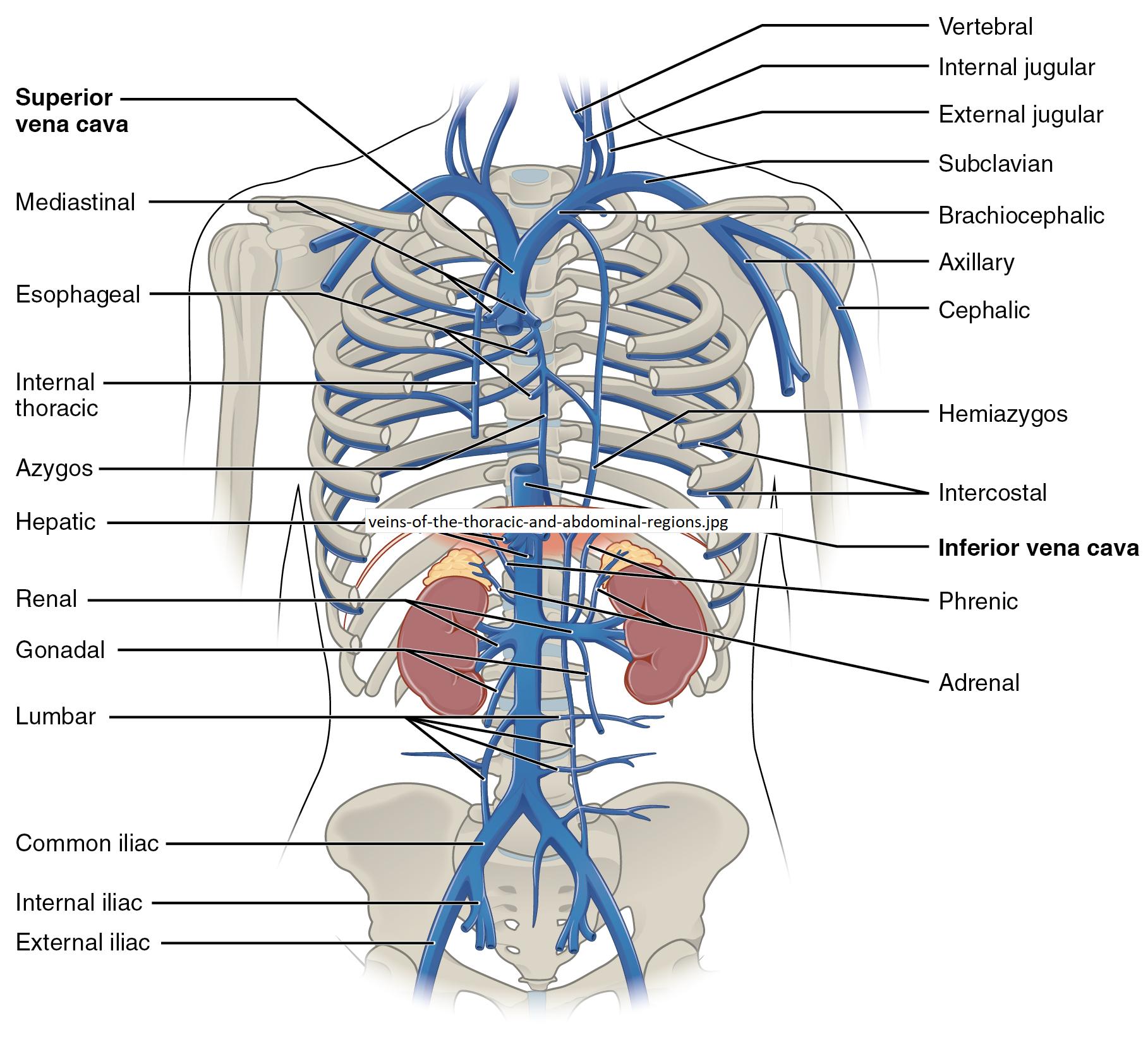
- Superior vena cava This large vein carries deoxygenated blood from the upper half of the body to the heart. It is formed by the junction of the brachiocephalic veins and plays a vital role in venous return.
- Mediastinal These veins drain blood from the mediastinum, the central compartment of the thoracic cavity. They contribute to the overall venous drainage by collecting blood from surrounding structures like the thymus and heart.
- Esophageal Located near the esophagus, these veins drain blood from the esophageal wall. They are essential for managing blood flow in the digestive tract region.
- Internal thoracic These veins run along the inner thoracic wall, draining blood from the chest wall and breast tissue. They connect to the brachiocephalic vein, aiding in upper body circulation.
- Azygos The azygos vein runs along the right side of the vertebral column, serving as an important collateral pathway. It drains blood from the thoracic wall and connects to the superior vena cava.
- Hepatic These veins drain blood from the liver, carrying nutrients and toxins processed by this organ. They empty into the inferior vena cava, facilitating liver function.
- Renal The renal veins drain blood from the kidneys, filtering waste and excess fluids. They play a crucial role in maintaining renal function and fluid balance.
- Gonadal These veins drain blood from the gonads (testes in males, ovaries in females). They extend downward to connect with the inferior vena cava or renal veins.
- Lumbar Located along the lumbar region, these veins drain blood from the back and abdominal wall muscles. They provide an essential drainage route for the lower spine area.
- Common iliac These veins form by the union of internal and external iliac veins, draining the pelvis and lower limbs. They are critical for lower body venous return.
- Internal iliac Draining the pelvic organs, these veins are vital for blood flow from the internal pelvic region. They merge to form the common iliac vein.
- External iliac These veins drain blood from the lower limbs and external pelvic region. They transition into the common iliac veins, supporting leg circulation.
- Vertebral Running along the vertebral column, these veins drain blood from the spinal cord and surrounding structures. They provide an alternative drainage pathway in the thoracic region.
- Internal jugular This vein drains blood from the brain, face, and neck, merging with the subclavian vein to form the brachiocephalic vein. It is essential for cranial circulation.
- External jugular Draining the exterior parts of the head and neck, this vein is visible superficially. It empties into the subclavian vein, aiding in upper body drainage.
- Subclavian This vein drains blood from the upper limbs and joins the internal jugular to form the brachiocephalic vein. It is key for arm and shoulder circulation.
- Brachiocephalic Formed by the union of the subclavian and internal jugular veins, it drains the head, neck, and upper limbs. It feeds into the superior vena cava.
- Axillary Draining the armpit and upper arm, this vein continues as the subclavian vein. It supports blood flow from the upper extremity.
- Cephalic Running along the lateral aspect of the arm, this vein drains the upper limb. It eventually joins the axillary vein, aiding arm circulation.
- Hemiarzygos A smaller vein on the left side of the vertebral column, it mirrors the azygos vein. It drains the left thoracic wall and connects to the azygos system.
- Intercostal These veins drain the intercostal spaces between the ribs. They contribute to thoracic wall drainage, connecting to the azygos and hemiazygos veins.
- Inferior vena cava This large vein returns deoxygenated blood from the lower body to the heart. It collects blood from the hepatic, renal, and iliac veins, playing a central role in circulation.
- Phrenic Draining the diaphragm, these veins are crucial for respiratory muscle blood flow. They empty into the inferior vena cava, supporting diaphragmatic function.
- Adrenal These veins drain the adrenal glands, which produce hormones like cortisol and aldosterone. They connect to the inferior vena cava, aiding endocrine regulation.
Anatomical Overview of the Veins
The veins of the thoracic and abdominal regions form a complex network essential for returning deoxygenated blood to the heart. This system ensures that blood from the upper body, including the head, neck, and arms, is efficiently transported via the superior vena cava, while the lower body relies on the inferior vena cava. The image highlights the anatomical layout, showing how these veins interconnect with various organs and tissues.
- The superior vena cava receives blood from tributaries like the brachiocephalic and azygos veins, reflecting its role in upper body drainage.
- The inferior vena cava collects blood from the hepatic, renal, and gonadal veins, showcasing its importance in lower body circulation.
- Smaller veins, such as the intercostal and lumbar, provide localized drainage, ensuring comprehensive coverage of the thoracic and abdominal walls.
Functional Significance in Circulation
The venous system depicted plays a pivotal role in maintaining hemodynamic stability. Each vein contributes to the overall process by collecting blood from specific regions and channeling it toward the heart.
- The renal veins filter blood to remove waste, supporting kidney health and fluid balance.
- The hepatic veins transport blood processed by the liver, ensuring detoxification and nutrient distribution.
- The internal jugular and external jugular veins are critical for draining the brain and face, preventing intracranial pressure buildup.
This interconnected network allows for collateral circulation, where alternative routes like the azygos and hemiazygos veins can compensate if primary pathways are obstructed. Such adaptability is vital for sustaining life during vascular challenges.
Clinical Relevance and Physical Examination
Understanding the anatomy of these veins is crucial for clinical assessments. Physical examination often involves palpating or visualizing these veins to assess circulatory health.
- The cephalic and axillary veins are accessible for venipuncture, aiding in diagnostic procedures.
- Swelling or distension of the superior vena cava may indicate obstruction, a condition requiring immediate attention.
- The phrenic veins’ proximity to the diaphragm makes them relevant in respiratory-related evaluations.
Healthcare professionals use this knowledge to diagnose conditions like thrombosis or compression syndromes, emphasizing the importance of anatomical awareness.
The veins of the thoracic and abdominal regions, as illustrated, are integral to the human circulatory system. Their precise anatomical arrangement ensures efficient blood return to the heart, supporting vital organ function and overall health. Exploring this network provides valuable insights into human physiology, underscoring the importance of each vein in maintaining homeostasis. Whether for educational purposes or clinical practice, a deep understanding of these structures enhances appreciation of the body’s intricate design.

