Explore the distinct anatomical features and functions of the male and female urethras, vital components of the urinary and reproductive systems. This article details how each urethra transports urine from the bladder, highlighting key differences relevant to health and medical conditions.
The Urethra: A Vital Conduit for Excretion
The urethra is a crucial tubular structure that serves as the final pathway for urine to exit the body from the urinary bladder. While its primary function is universal—to transport urine—its anatomical configuration differs significantly between males and females. These anatomical distinctions have important implications for urinary health, susceptibility to certain medical conditions, and reproductive functions.
Understanding the specific characteristics of the male and female urethras is fundamental in urology and reproductive health. This knowledge helps explain common conditions like urinary tract infections (UTIs) and aids in the diagnosis and treatment of various genitourinary disorders.
Key aspects of the urethra include:
- Transporting urine from the bladder to the outside of the body.
- Significant anatomical differences between sexes.
- Dual function in males (urinary and reproductive).
These characteristics highlight its integral role in both excretion and reproduction.
Female Urethra: Anatomy and Associated Structures
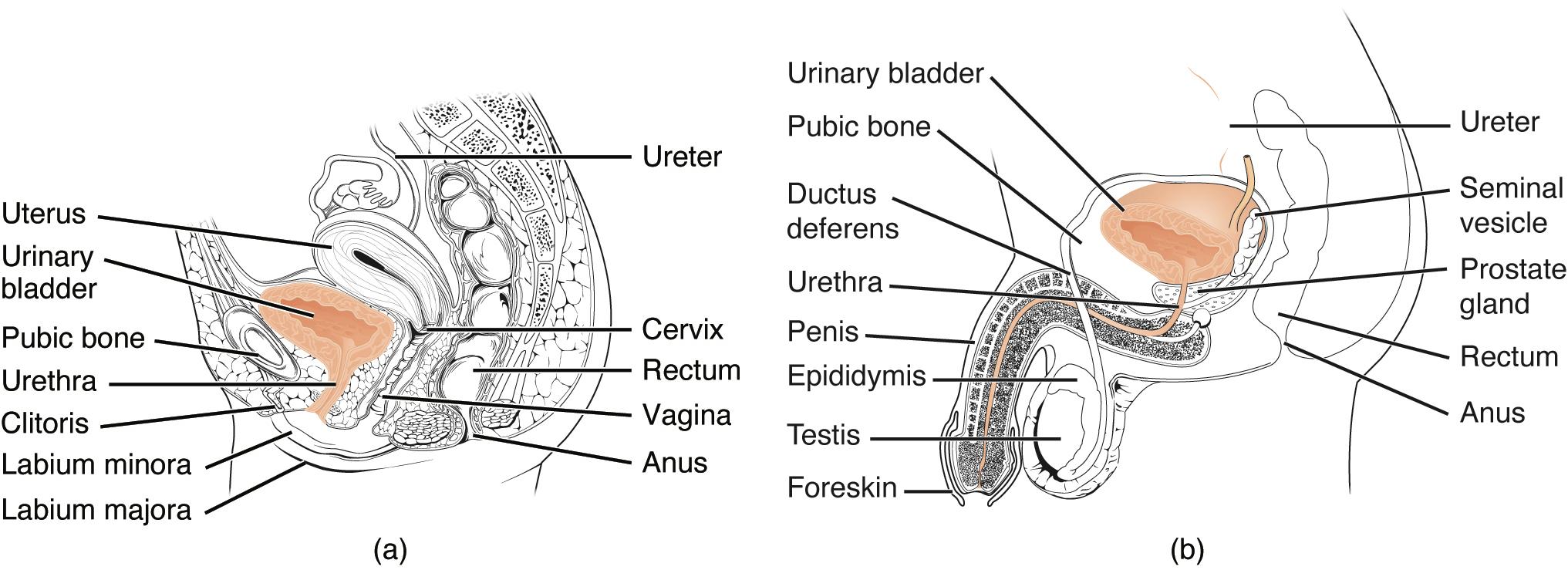
(a) Female Urethra: In females, the urethra is a relatively short tube, typically about 3-4 centimeters (1.5 inches) long, extending from the urinary bladder to the outside of the body. Its shorter length makes females more susceptible to urinary tract infections compared to males.
Ureter: These are tubes that transport urine from the kidneys to the urinary bladder. There are two ureters, one from each kidney, descending into the pelvis.
Uterus: A pear-shaped muscular organ in the female reproductive system where a fertilized egg implants and develops during pregnancy. It is located between the bladder and the rectum.
Urinary bladder: A hollow, muscular organ that stores urine received from the ureters until it is voluntarily expelled from the body through the urethra.
Pubic bone: A bone located at the front of the pelvis, forming the anterior part of the pelvic girdle. It provides protection and structural support to the urinary and reproductive organs.
Urethra (female): As described, this short tube carries urine from the bladder to the exterior. Its external opening, the external urethral orifice, is located between the clitoris and the vaginal opening.
Clitoris: A highly sensitive erectile organ in the female external genitalia, playing a key role in sexual arousal. It is located anterior to the urethral opening.
Labium minora: Two small folds of skin located on either side of the vaginal opening, medial to the labia majora. They protect the clitoris and urethral and vaginal openings.
Labium majora: Two larger, fleshy folds of skin that enclose the labia minora and other external female genital organs. They provide protection and contribute to the external appearance of the vulva.
Cervix: The lower, narrow part of the uterus that forms a canal connecting the uterus to the vagina. It acts as a gateway between the uterus and the vaginal canal.
Rectum: The final section of the large intestine, terminating at the anus. It stores feces before defecation and is located posterior to the vagina and uterus in females.
Vagina: A muscular, elastic tube that connects the uterus to the outside of the body. It serves as the birth canal, receives the penis during sexual intercourse, and is the pathway for menstrual flow.
Anus: The external opening of the rectum, through which feces are expelled from the body.
Male Urethra: Anatomy and Associated Structures
(b) Male Urethra: In males, the urethra is significantly longer, typically about 18-20 centimeters (7-8 inches), and extends from the urinary bladder through the penis to the outside. It serves a dual function, transporting both urine and semen.
Ureter: Similar to females, these tubes transport urine from the kidneys to the urinary bladder.
Urinary bladder: A muscular sac that stores urine before its expulsion, identical in function to the female bladder but located slightly differently due to surrounding reproductive organs.
Pubic bone: The anterior pelvic bone, providing structural support and protection to the male genitourinary organs.
Ductus deferens (Vas deferens): A muscular tube that transports sperm from the epididymis to the ejaculatory duct. It loops over the ureter and runs behind the bladder.
Seminal vesicle: Glands located behind the urinary bladder that produce a fluid component of semen, rich in fructose to nourish sperm.
Prostate gland: A gland located below the urinary bladder and surrounding the urethra. It secretes a fluid that contributes to semen volume and helps activate sperm.
Rectum: The final section of the large intestine, located posterior to the bladder and prostate in males.
Urethra (male): This longer tube carries both urine and semen. It is divided into three main sections: prostatic, membranous, and spongy (penile) urethra.
Penis: The male external sexual organ, containing the urethra and serving both urinary and reproductive functions.
Epididymis: A coiled tube located on the posterior side of each testis, where sperm mature and are stored.
Testis: The primary male reproductive organs, producing sperm and male hormones (testosterone).
Foreskin: A retractable fold of skin that covers the tip of the penis (glans penis) in uncircumcised males.
Anus: The external opening of the rectum, for the expulsion of feces.
Functional Differences and Clinical Relevance
The most notable difference between the male and female urethra is its length and dual function in males. The female urethra is considerably shorter and serves only the urinary system, meaning it transports only urine. Its brevity and close proximity to the anus make it more susceptible to bacterial contamination and, consequently, a higher incidence of urinary tract infections (UTIs) in females. Bacteria can more easily travel up the short urethra to the bladder.
In contrast, the male urethra is much longer and is a shared pathway for both the urinary and reproductive systems. It transports urine from the bladder and semen during ejaculation. This dual role means that diseases affecting the male urethra can impact both urinary and reproductive functions. Conditions such as benign prostatic hyperplasia (BPH), an enlargement of the prostate gland common in older men, can compress the prostatic urethra, leading to urinary flow obstruction and symptoms like difficulty urinating or frequent urination. Urethral strictures, which are narrowing of the urethra due to scar tissue, can also occur in both sexes but are more common in males due to trauma or infection.
Conclusion
The urethra, despite its seemingly simple role in urine excretion, exhibits significant anatomical and functional distinctions between males and females, which bear critical clinical implications. The female urethra’s shorter length predisposes women to a higher incidence of urinary tract infections, while the male urethra’s longer, dual-purpose pathway can be affected by prostate issues and strictures. A comprehensive understanding of these differences is indispensable for healthcare professionals in diagnosing, treating, and preventing various genitourinary conditions, underscoring the importance of sex-specific anatomical knowledge in clinical practice.

