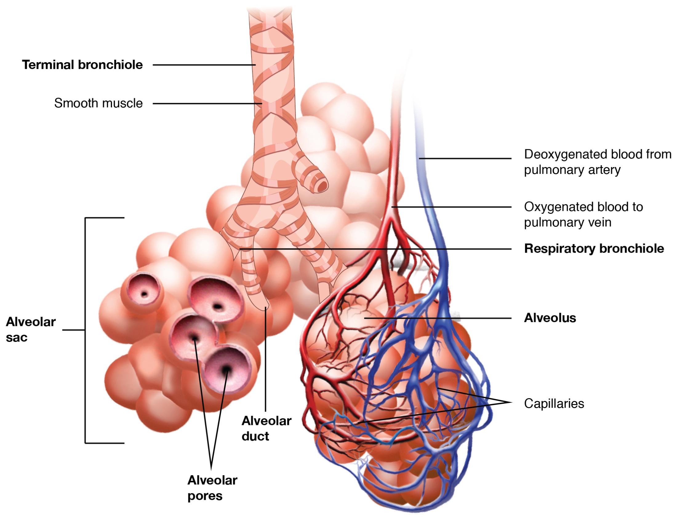
The respiratory zone represents the final stage of the respiratory system where gas exchange occurs, beginning with bronchioles leading to alveolar sacs. This critical region, nestled deep within the lungs, facilitates the transfer of oxygen into the bloodstream and the expulsion of carbon dioxide, sustaining life with every breath. Exploring this anatomical diagram provides a clear view of the structures involved, offering valuable insights into the intricate process of respiration.
Key Anatomical Labels in the Diagram
This section breaks down each labeled component, highlighting their roles and positions within the respiratory zone.
Bronchiole: The bronchiole is a small airway branching from the bronchi, delivering air to the alveolar sacs. Its thin walls and lack of cartilage allow for greater flexibility and adjustability during breathing.
Alveolar sac: The alveolar sac is a cluster of alveoli where gas exchange takes place, surrounded by a network of capillaries. Its thin walls, composed of type I and type II pneumocytes, maximize oxygen and carbon dioxide diffusion.
Alveolus: The alveolus is a tiny air sac within the alveolar sac, serving as the primary site for oxygen to enter the blood and carbon dioxide to be removed. Its surface is lined with surfactant, reducing surface tension to prevent collapse.
Capillary: The capillary is a microscopic blood vessel surrounding the alveoli, enabling gas exchange through its thin endothelial walls. It carries deoxygenated blood to the alveoli and returns oxygenated blood to the pulmonary veins.
Type I pneumocyte: The type I pneumocyte is a flat, thin cell forming the majority of the alveolar wall, optimizing gas diffusion. It provides a minimal barrier for efficient oxygen and carbon dioxide exchange.
Type II pneumocyte: The type II pneumocyte is a cuboidal cell that produces and secretes surfactant, maintaining alveolar stability. It also plays a role in repairing the alveolar lining after injury.
Structure of the Respiratory Zone
The respiratory zone’s architecture centers around its air sacs and supporting structures. This design ensures effective gas exchange within the lungs.
- The bronchiole transitions from conductive to respiratory airways, lacking cartilage.
- Alveolar sacs house multiple alveoli, increasing the surface area for diffusion.
- The alveolus’s thin walls facilitate rapid gas transfer across membranes.
- Capillaries form a dense network, enhancing blood-gas interaction.
- This structure supports the lungs’ vast respiratory capacity.
Cellular Components and Their Functions
The cells within the respiratory zone are specialized for gas exchange and maintenance. These elements work together to sustain lung function.
- Type I pneumocytes provide a thin barrier, maximizing diffusion efficiency.
- Type II pneumocytes secrete surfactant, preventing alveolar collapse.
- Capillary endothelial cells allow oxygen to bind with hemoglobin.
- Alveolar macrophages patrol the surface, clearing debris and pathogens.
- This cellular synergy ensures continuous respiratory health.
Physiological Role in Gas Exchange
The respiratory zone is where oxygen and carbon dioxide exchange occurs seamlessly. Its anatomy supports the body’s metabolic needs.
- The alveolus’s large surface area, about 70 square meters, enhances gas transfer.
- Capillaries facilitate oxygen binding to red blood cells for circulation.
- Surfactant reduces surface tension, keeping alveoli open during exhalation.
- The bronchiole delivers air, adjusting to breathing rate changes.
- This process maintains blood pH through efficient carbon dioxide removal.
Clinical Relevance and Anatomical Variations
Understanding the respiratory zone’s anatomy aids in diagnosing and treating lung conditions. Variations can influence clinical outcomes.
- Alveolar damage, as in pulmonary edema, impairs gas exchange.
- Bronchiole constriction in asthma reduces airflow to alveoli.
- Type II pneumocyte dysfunction can lead to surfactant deficiency, causing collapse.
- Capillary leakage in acute respiratory distress syndrome affects oxygenation.
- Imaging like CT scans assesses these structures for therapeutic planning.
The respiratory zone’s intricate design, from bronchioles to alveolar sacs, underscores its vital role in gas exchange and oxygenation. By studying this anatomical diagram, one gains a profound appreciation for the lungs’ ability to sustain life, revealing the elegance of this essential respiratory system.

