The phospholipid bilayer is a critical structure in biology, forming the foundation of all cell membranes. This dynamic and essential component regulates what enters and exits the cell, maintaining its internal environment while facilitating communication with the extracellular space. Explore the intricate details of its structure and function through this detailed analysis, ideal for those seeking a deeper understanding of cellular biology.
Labels Introduction
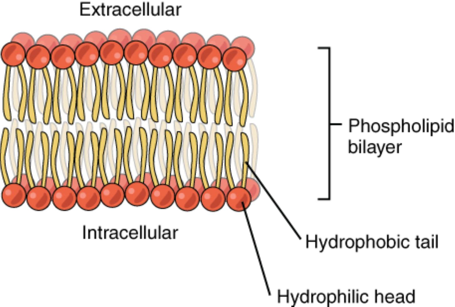
Extracellular
The extracellular region represents the outer environment surrounding the cell, where the hydrophilic heads of the phospholipids are exposed. This layer interacts with the external fluid, playing a key role in cell signaling and nutrient exchange.
Intracellular
The intracellular area is the inner environment of the cell, also featuring hydrophilic heads that face the cytoplasm. This side is crucial for maintaining internal homeostasis and supporting cellular processes.
Phospholipid bilayer
The phospholipid bilayer is composed of two layers of phospholipids arranged tail to tail, creating a semi-permeable barrier. This structure provides stability and flexibility, essential for the membrane’s protective and regulatory functions.
Hydrophobic tail
The hydrophobic tail, consisting of fatty acid chains, repels water and forms the interior of the bilayer. This nonpolar region prevents the passage of water-soluble substances, contributing to the membrane’s selective permeability.
Hydrophilic head
The hydrophilic head, containing a phosphate group, attracts water and is oriented toward the extracellular and intracellular fluids. This polar region enables interactions with the aqueous environments on both sides of the membrane.
Structural Overview of the Phospholipid Bilayer
The phospholipid bilayer serves as the basic framework of cell membranes, a vital component in all living organisms. Comprised of two layers of phospholipids, each molecule features a hydrophilic head and a hydrophobic tail, arranged to create a stable yet fluid structure. This organization allows the membrane to protect the cell while permitting essential exchanges.
- The bilayer’s unique arrangement stems from the amphipathic nature of phospholipids, which have both hydrophilic and hydrophobic properties.
- This structure is dynamic, allowing proteins and other molecules to embed within it, enhancing its functionality.
Role in Cellular Function
The phospholipid bilayer plays a pivotal role in maintaining cellular integrity and function. By forming a barrier, it controls the movement of substances, ensuring the cell’s internal environment remains stable. Additionally, it hosts various proteins that facilitate transport, signaling, and enzymatic activities.
- Selective permeability is a key attribute, allowing small or lipid-soluble molecules to pass while blocking larger or charged ones.
- Embedded proteins, such as ion channels and receptors, enable communication and transport across the membrane.
Chemical Composition and Properties
Phospholipids are the primary building blocks of the bilayer, consisting of a glycerol backbone, two fatty acid tails, and a phosphate group. The hydrophobic tails, typically nonpolar, associate with each other to form a water-repellent core. In contrast, the hydrophilic heads interact with the aqueous environments on either side.
- The fatty acid tails can vary in length and saturation, influencing the membrane’s fluidity and flexibility.
- The phosphate group, often linked to additional molecules like choline or serine, contributes to the head’s polarity.
Interaction with the Extracellular and Intracellular Environment
The extracellular and intracellular regions define the bilayer’s boundaries, with hydrophilic heads facing these aqueous spaces. This orientation ensures the membrane can interact with external signals and internal cellular components. The stability of this interaction is crucial for processes like endocytosis and exocytosis.
- The extracellular surface may contain glycolipids and glycoproteins, aiding in cell recognition and adhesion.
- Inside the cell, the intracellular side supports the attachment of the cytoskeleton, maintaining cell shape.
Importance in Health and Disease Prevention
While the image does not depict specific diseases, the phospholipid bilayer’s integrity is vital for preventing cellular damage. Disruptions in its structure can lead to conditions like membrane instability or impaired transport. Understanding its normal function provides insight into maintaining cellular health.
- A balanced lipid composition helps resist oxidative stress and inflammation.
- Abnormalities in bilayer fluidity are linked to various pathological states, emphasizing its role in homeostasis.
Advanced Biological Insights
For those delving deeper, the phospholipid bilayer’s role extends to supporting specialized functions in different cell types. For instance, in nerve cells, the bilayer facilitates rapid signal transmission through ion channel regulation. In endocrine cells, it supports hormone release, such as thyroid hormones T3 and T4, which regulate metabolism.
- The bilayer’s fluidity adapts to temperature changes, ensuring functionality across environments.
- Cholesterol molecules within the bilayer modulate its rigidity, balancing fluidity and stability.
Conclusion
The phospholipid bilayer is a remarkable structure that underpins the functionality of all cell membranes. Its unique composition and dynamic properties enable it to serve as a protective barrier, a selective gate, and a platform for critical cellular processes. By exploring its components and roles, we gain a greater appreciation for the complexity of cellular biology and the importance of maintaining membrane health.

