Explore the intricate pharynx anatomical structure with this detailed diagram, illustrating its role as a vital passageway from the nostrils to the esophagus and larynx. Learn about the nasopharynx, oropharynx, and laryngopharynx, and their critical functions in both respiration and digestion.
The pharynx, commonly known as the throat, is a musculomembranous tube that serves as a crucial anatomical crossroads for both the respiratory and digestive systems. Extending from the base of the skull to the level of the sixth cervical vertebra, it facilitates the passage of air to the lungs and food to the esophagus. Understanding its segmented structure—the nasopharynx, oropharynx, and laryngopharynx—is essential for comprehending the complex processes of breathing, swallowing, and speech.
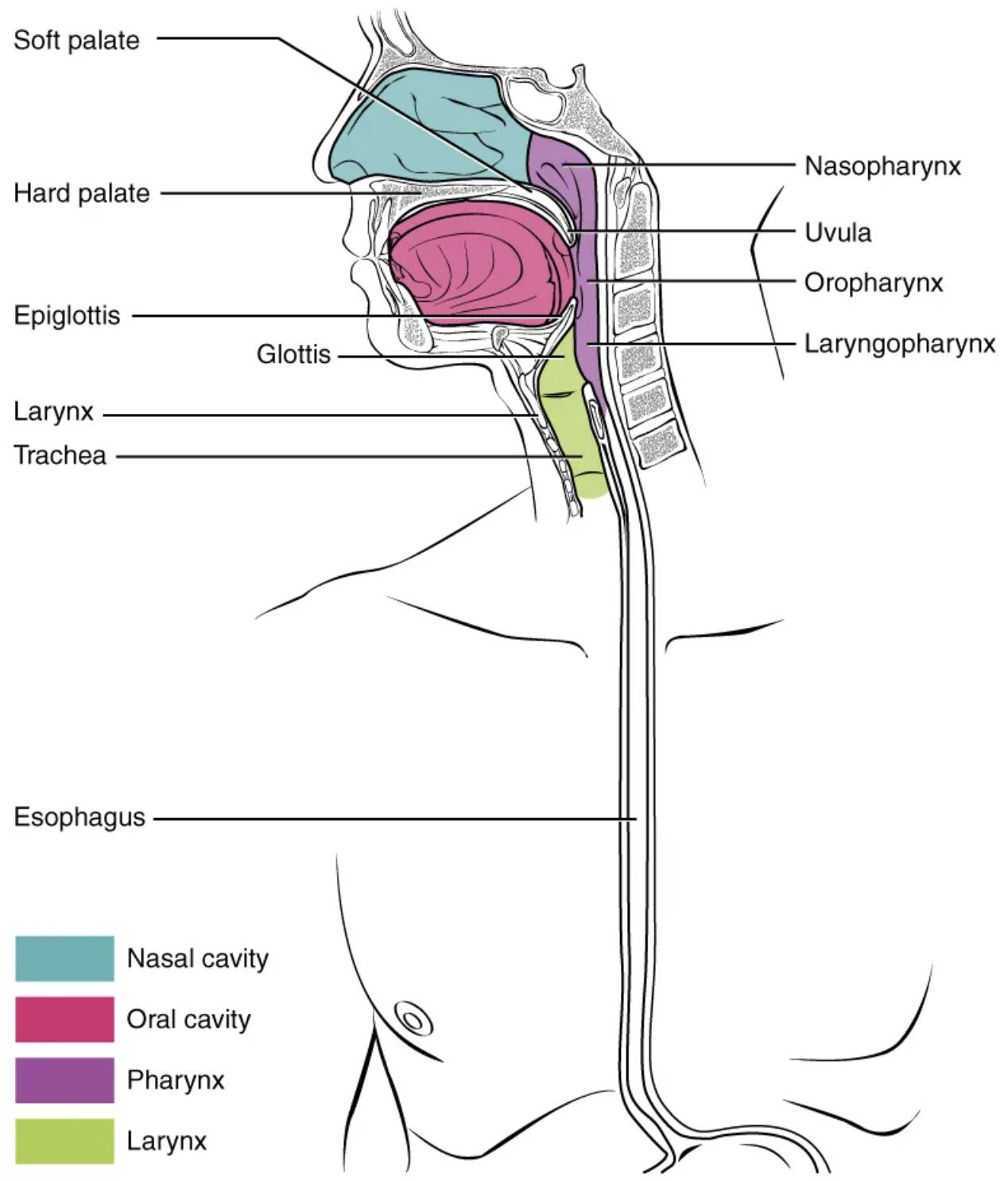
Soft palate: The soft palate is the posterior, fleshy part of the roof of the mouth, composed of muscle and connective tissue. During swallowing, it elevates to seal off the nasopharynx, preventing food from entering the nasal cavity.
Hard palate: The hard palate forms the anterior, bony part of the roof of the mouth, separating the oral cavity from the nasal cavity. It provides a rigid surface against which the tongue can press food during mastication and swallowing.
Epiglottis: The epiglottis is a leaf-shaped flap of cartilage located at the base of the tongue, positioned superior to the glottis. Its primary function is to fold down over the glottis during swallowing, directing food and liquids away from the trachea and into the esophagus.
Glottis: The glottis refers to the vocal folds (true vocal cords) and the opening between them, located within the larynx. It is crucial for voice production and plays a role in preventing food from entering the lower respiratory tract.
Larynx: The larynx, or voice box, is a cartilaginous structure located in the anterior neck, inferior to the pharynx. It houses the vocal cords, enabling speech, and also serves as a protective passageway for air into the trachea.
Trachea: The trachea, commonly known as the windpipe, is a cartilaginous tube extending from the larynx into the chest cavity. Its main function is to provide a clear airway for air to enter and exit the lungs.
Esophagus: The esophagus is a muscular tube that connects the pharynx to the stomach. Its primary role is to transport food from the pharynx to the stomach through wave-like muscular contractions called peristalsis.
Nasopharynx: The nasopharynx is the uppermost part of the pharynx, located posterior to the nasal cavity and extending to the soft palate. It serves exclusively as an air passageway and houses the pharyngeal tonsils.
Uvula: The uvula is a small, fleshy projection hanging from the middle of the soft palate. It plays a role in speech articulation, particularly in forming certain sounds, and helps prevent food from entering the nasopharynx during swallowing.
Oropharynx: The oropharynx is the middle part of the pharynx, located posterior to the oral cavity and extending from the soft palate to the epiglottis. It serves as a common passageway for both air and food, and houses the palatine and lingual tonsils.
Laryngopharynx: The laryngopharynx is the lowest part of the pharynx, extending from the epiglottis to the esophagus. It also serves as a common passageway for both air and food, directing air into the larynx and food into the esophagus.
The Pharynx: A Critical Anatomical Junction
The pharynx, a cone-shaped muscular tube approximately 12-14 cm long, is an anatomical region of immense functional importance in the human body. It acts as a shared conduit, uniquely designed to manage the passage of both air for respiration and food for digestion. Its strategic position, extending from the nasal cavity and mouth down to the larynx and esophagus, necessitates a complex coordination of muscular contractions and structural adjustments to ensure that substances are directed to their correct pathways, preventing aspiration and facilitating vital bodily functions.
The pharynx is anatomically divided into three distinct regions, each with specialized roles:
-
Nasopharynx: The superior portion, located behind the nasal cavity, is solely dedicated to air passage. It connects to the Eustachian tubes, helping to equalize pressure in the middle ear.
-
Oropharynx: The middle portion, behind the oral cavity, serves as a common pathway for both air and food. This is where the gag reflex is initiated.
-
Laryngopharynx: The inferior portion, extending from the epiglottis to the esophagus, also handles both air and food. This segment is crucial in directing food into the esophagus and air into the larynx.
The intricate coordination of structures within and around the pharynx, particularly during the act of swallowing (deglutition), is remarkable. As food is moved from the oral cavity into the oropharynx, the soft palate and uvula elevate to seal off the nasopharynx, preventing food from entering the nasal passages. Simultaneously, the larynx rises, and the epiglottis folds downward, covering the glottis and effectively preventing food from entering the trachea and subsequently the lungs. This precise mechanism ensures that food is channeled exclusively into the esophagus, while air can continue its unobstructed path to the lungs. Any disruption to this delicate balance, perhaps due to neurological conditions or structural abnormalities, can lead to dysphagia (difficulty swallowing) or a heightened risk of aspiration, highlighting the pharynx’s critical role in maintaining both respiratory and digestive integrity.
In conclusion, the pharynx stands as a vital junction in human anatomy, orchestrating the complex interplay between breathing and eating. Its segmented structure and the coordinated actions of surrounding components are essential for directing air and food appropriately, protecting the airway, and enabling clear speech. A comprehensive understanding of the pharynx’s structure and function is paramount for medical professionals in fields such as otolaryngology, gastroenterology, and speech pathology, as it underpins the assessment and treatment of a wide range of respiratory and digestive disorders.

