The sense of smell, a vital component of human perception, begins in the nasal cavity where the olfactory system processes airborne molecules. This intricate system, as depicted in the image, involves specialized structures within the olfactory epithelium and connections to the brain, enabling the detection and interpretation of odors. This article explores the anatomy and physiology of these components, providing a detailed look at how the olfactory system functions to enhance our sensory experience.
Labeled Parts of the Olfactory System
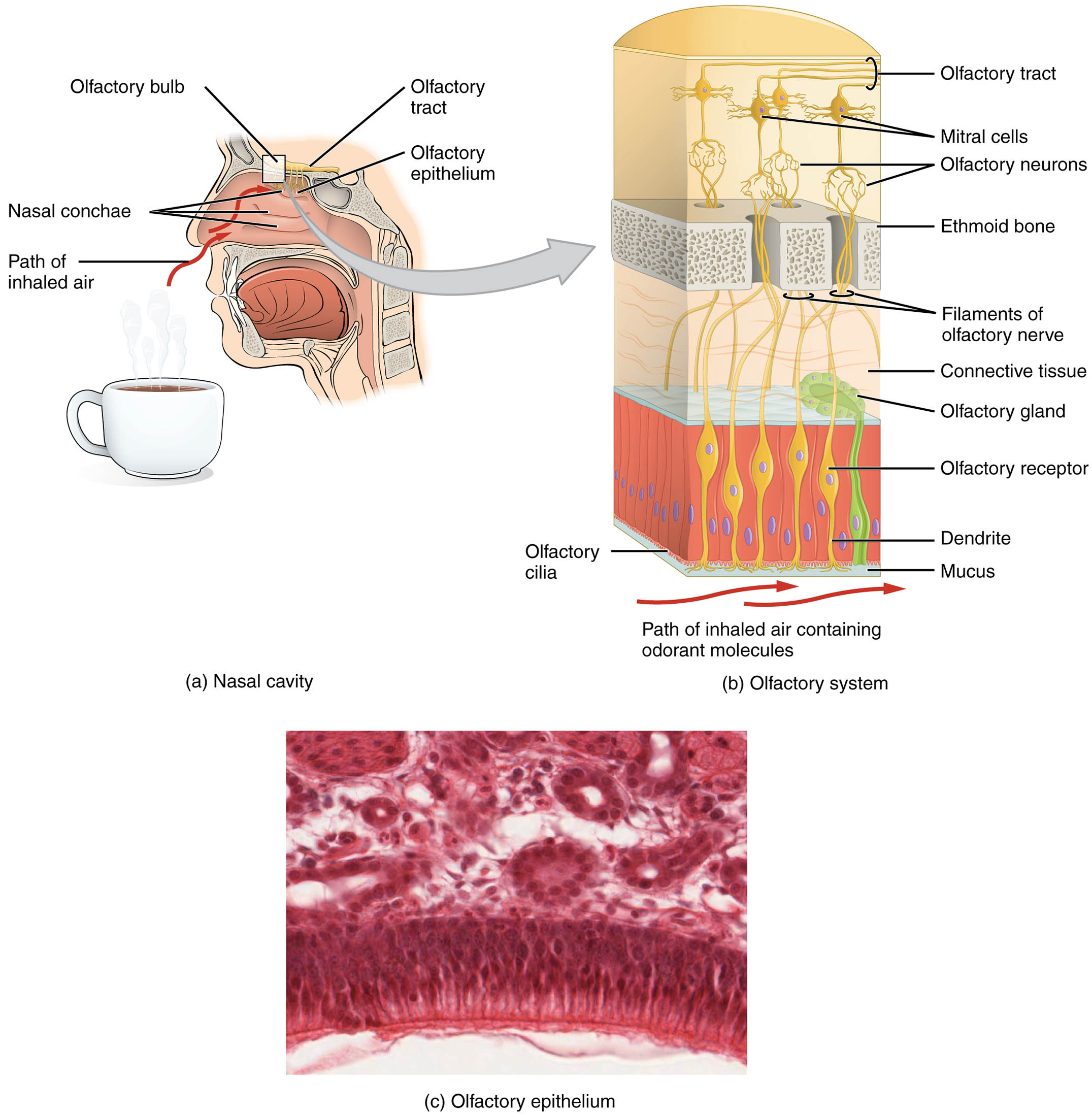
Olfactory bulb The olfactory bulb is a neural structure located at the base of the brain, where axons from olfactory receptor neurons synapse with mitral cells. It processes incoming odor information and relays it to higher brain regions, such as the olfactory tract, for further interpretation.
Olfactory tract The olfactory tract is a bundle of nerve fibers that extends from the olfactory bulb to the olfactory cortex, transmitting processed smell signals. It plays a crucial role in connecting the peripheral olfactory system to the central nervous system for conscious perception.
Olfactory epithelium The olfactory epithelium is a specialized tissue lining the nasal cavity, containing olfactory receptor neurons and supporting cells. It serves as the primary site for detecting odorant molecules, initiating the sensory process of smell.
Mitral cells Mitral cells are large neurons within the olfactory bulb that receive input from olfactory receptor neurons and integrate odor information. They project axons along the olfactory tract, amplifying and refining the smell signal before it reaches the brain.
Olfactory neurons Olfactory neurons are bipolar sensory cells within the olfactory epithelium, equipped with dendrites that detect odorants. Their axons form the filaments of the olfactory nerve, transmitting sensory data to the olfactory bulb.
Ethmoid bone The ethmoid bone is a cranial bone with a cribriform plate that allows olfactory nerve filaments to pass from the nasal cavity to the brain. It provides structural support and protection to the delicate olfactory pathways.
Filaments of olfactory nerve The filaments of the olfactory nerve are the axons of olfactory neurons that penetrate the ethmoid bone to synapse in the olfactory bulb. They form the first cranial nerve (CN I), essential for conveying olfactory signals to the brain.
Connective tissue Connective tissue underlies the olfactory epithelium, providing structural support and housing olfactory glands. It helps maintain the integrity of the epithelium and facilitates the secretion of mucus.
Olfactory gland The olfactory gland, located within the connective tissue, produces mucus that moistens the olfactory epithelium. This mucus traps odorant molecules, enabling receptor cells to detect and process them effectively.
Olfactory receptor The olfactory receptor is a specialized protein on the dendrite of olfactory neurons that binds to odorant molecules. This binding triggers a signal transduction process, initiating the perception of smell.
Dendrite The dendrite is the branched extension of an olfactory neuron that contains olfactory receptors and cilia, detecting odorant molecules in the mucus. It converts chemical stimuli into electrical impulses for neural transmission.
Cilia Cilia are hair-like projections on the dendrites of olfactory neurons, increasing the surface area for odorant detection. They move within the mucus layer, enhancing the efficiency of smell perception.
Mucus Mucus is a protective and moist layer secreted by olfactory glands, covering the olfactory epithelium. It dissolves odorant molecules, allowing them to interact with olfactory receptors for sensory processing.
Nasal conchae The nasal conchae are bony structures within the nasal cavity that increase air turbulence and surface area. They help direct inhaled air toward the olfactory epithelium, optimizing odor detection.
Path of inhaled air The path of inhaled air travels through the nasal cavity, passing over the nasal conchae and reaching the olfactory epithelium. This airflow carries odorant molecules to the receptors, initiating the olfactory process.
Anatomical Overview of the Olfactory System
The olfactory system is a sophisticated sensory mechanism that begins in the nasal cavity and extends to the brain. Its anatomical design ensures efficient detection and processing of odors, integrating with other sensory pathways.
- Nasal cavity role: The nasal conchae enhance air circulation, guiding inhaled air containing odorant molecules to the olfactory epithelium.
- Epithelial structure: The olfactory epithelium houses receptor cells, supported by connective tissue and glands, forming a robust sensory layer.
- Neural pathway: Olfactory neurons send axons through the ethmoid bone, forming the olfactory nerve filaments that connect to the olfactory bulb.
- Bulb processing: Mitral cells within the olfactory bulb refine sensory input, sending signals via the olfactory tract to the brain for interpretation.
- Microscopic view: The micrograph of the olfactory epithelium reveals the dense arrangement of receptor cells, highlighting its cellular complexity.
Physiological Functions of the Olfactory System
The olfactory system converts chemical stimuli into neural signals, playing a key role in smell perception and related functions. Its physiological processes are finely tuned to environmental cues.
- Odorant detection: Olfactory receptors on dendrites bind to odorant molecules dissolved in mucus, triggering a cascade of electrical signals.
- Signal transduction: Cilia amplify the receptor response, while olfactory neurons convert chemical stimuli into action potentials.
- Neural transmission: Axons of olfactory neurons, bundled as filaments, transmit signals through the ethmoid bone to the olfactory bulb.
- Integration: Mitral cells process and relay refined odor information along the olfactory tract to the olfactory cortex and limbic system.
- Protective mechanism: Mucus and nasal conchae filter and humidify air, protecting the epithelium while facilitating odorant access.
Developmental and Cellular Dynamics
The olfactory system develops early in fetal life, with its structures maturing to support smell perception by birth. Cellular turnover and adaptation ensure its functionality throughout life.
- Embryonic formation: The olfactory epithelium arises from ectodermal tissue, with neurons and glands differentiating during gestation.
- Cellular renewal: Olfactory neurons are regularly replaced by stem cells within the epithelium, maintaining sensory capability.
- Nerve growth: Axons extend through the ethmoid bone, forming the olfactory nerve, with synapses established in the bulb prenatally.
- Evolutionary traits: The system’s design reflects adaptations for detecting food and predators, conserved across species.
- Age-related changes: Sensory decline with age may reduce cilia and receptor efficiency, affecting smell acuity.
Clinical Relevance and Olfactory Disorders
Understanding the olfactory system aids in diagnosing and managing smell-related conditions. Clinical assessments often focus on epithelial and neural integrity to identify dysfunction.
- Anosmia: Loss of smell can result from damage to olfactory neurons or the epithelium, often due to head trauma or viral infections.
- Hyposmia: Reduced smell sensitivity may indicate aging or neurodegenerative diseases like Parkinson’s, affecting quality of life.
- Diagnostic tools: Olfactory testing with odor identification kits and imaging of the ethmoid bone assess system health.
- Therapeutic options: Treatments include addressing underlying causes, such as sinus surgery or olfactory training, to restore function.
- Associated risks: Loss of smell can impact nutrition and safety, underscoring the need for early intervention.
In conclusion, the olfactory system’s anatomy, from the nasal cavity to the olfactory bulb, showcases a remarkable integration of structure and function. This sensory pathway not only enriches our experience with scents but also serves as a diagnostic window into neurological and respiratory health, making its study essential for comprehensive care.

