The maculae of the utricle and saccule play a crucial role in our sense of balance by detecting linear accelerations, including the pull of gravity and straight-line movements of the head. This intricate system within the inner ear helps maintain equilibrium and spatial orientation, making it essential for everyday activities like walking or tilting the head.
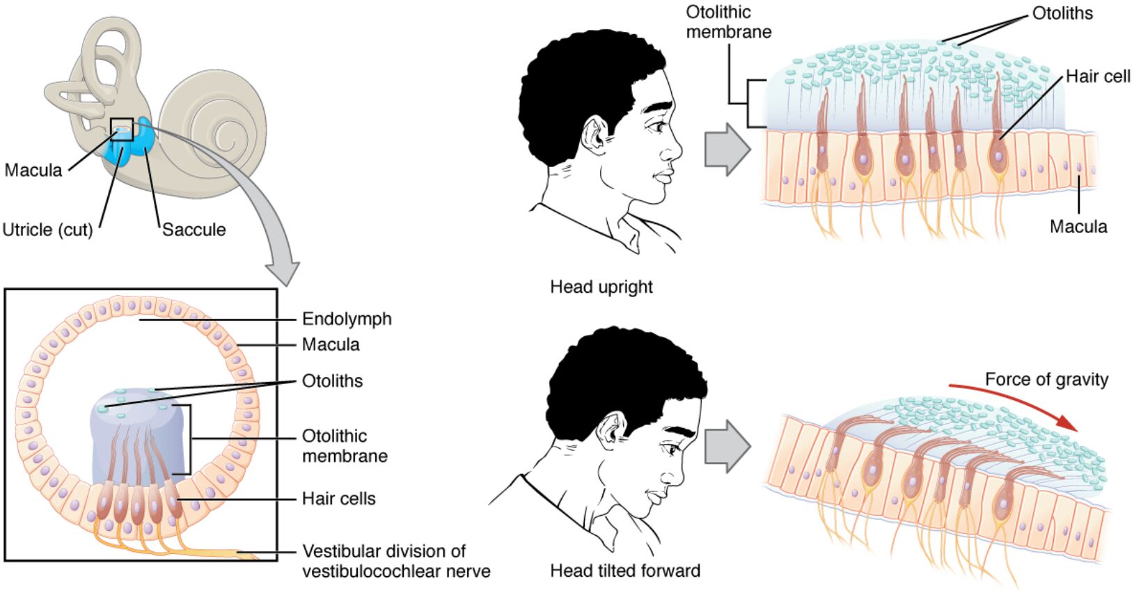
Macula The macula is a sensory patch located in the utricle and saccule of the inner ear, consisting of hair cells embedded in a gelatinous matrix. It detects changes in head position relative to gravity by responding to the movement of otoliths, which shear against the hair cell stereocilia.
Utricle (cut) The utricle is a pouch-like structure in the vestibular system, shown here in a cut view to reveal its internal components. It primarily senses horizontal linear accelerations, such as those experienced during forward or sideways movements.
Saccule The saccule is another vestibular sac adjacent to the utricle, oriented vertically to detect up-and-down accelerations. It works in tandem with the utricle to provide comprehensive information about the body’s position in space.
Otolithic membrane The otolithic membrane is a gelatinous layer overlying the hair cells in the macula, embedded with calcium carbonate crystals called otoliths. This membrane shifts in response to linear forces, bending the underlying stereocilia to generate neural signals.
Otoliths Otoliths are small, dense crystals made of calcium carbonate that rest on the otolithic membrane. Their inertia causes them to lag behind or move differently from the surrounding structures during acceleration, triggering sensory responses in the hair cells.
Hair cell A hair cell is a specialized receptor cell in the macula with protruding stereocilia that detect mechanical deflections. These cells convert physical movements into electrical signals transmitted to the brain via the vestibular nerve.
Endolymph Endolymph is the potassium-rich fluid filling the inner ear’s membranous labyrinth, including the utricle and saccule. It provides the medium in which the maculae operate, allowing for the fluid dynamics that influence hair cell stimulation.
Hair cells Hair cells are mechanoreceptors that line the macula, featuring bundles of stereocilia and a kinocilium for directional sensitivity. They depolarize or hyperpolarize based on the direction of bending, encoding the specifics of linear acceleration.
Vestibular division of vestibulocochlear nerve The vestibular division of vestibulocochlear nerve carries sensory information from the maculae to the brainstem. This nerve branch is vital for processing balance signals, integrating them with visual and proprioceptive inputs for postural control.
Force of gravity The force of gravity acts as a constant linear acceleration detected by the maculae, particularly when the head tilts. It causes the otoliths to pull downward, bending hair cell stereocilia and signaling changes in head orientation.
Anatomy of the Vestibular System
The vestibular system, housed in the inner ear, is responsible for detecting motion and position to maintain balance. Key components like the utricle and saccule contain the maculae, which are ingeniously designed to sense linear changes.
- The utricle and saccule are fluid-filled sacs connected to the semicircular canals, forming the membranous labyrinth.
- Endolymph within these structures has a specific density that interacts with the otolithic membrane during movement.
- The macula in the utricle is oriented horizontally, while in the saccule it is vertical, allowing detection along different axes.
- Hair cells are arranged in a striola, a central dividing line where polarization reverses for bidirectional sensing.
- Supporting cells surround hair cells, providing structural integrity and aiding in ion homeostasis.
- The vestibulocochlear nerve, or cranial nerve VIII, has a vestibular branch that innervates these sensory epithelia.
Physiology of Linear Acceleration Detection
Linear acceleration, whether from gravity or motion, is transduced by the maculae through a mechanical process involving inertia. When the head moves, the difference in momentum between otoliths and the surrounding gel leads to shear forces.
- In a stationary upright position, otoliths exert a steady pull on stereocilia due to gravity, establishing a baseline signal.
- Tilting the head forward causes otoliths to slide, bending stereocilia toward or away from the kinocilium.
- Depolarization occurs when stereocilia bend toward the kinocilium, increasing neurotransmitter release to afferent nerves.
- Hyperpolarization happens in the opposite direction, reducing firing rates and providing contrast in sensory input.
- This push-pull mechanism allows the brain to interpret the direction and magnitude of acceleration.
- Integration in the vestibular nuclei processes these signals for reflexes like the vestibulo-ocular reflex.
Role of Otoliths and Otolithic Membrane
Otoliths serve as inertial masses that enhance the sensitivity of the maculae to low-frequency accelerations. The otolithic membrane acts as a coupling layer, transmitting forces efficiently to the hair cells below.
- Composed primarily of calcium carbonate in the form of aragonite crystals, otoliths vary in size from 1 to 30 micrometers.
- The membrane’s gelatinous matrix, rich in glycoproteins, allows for viscoelastic properties that dampen rapid movements.
- During linear acceleration, the membrane shears relative to the macular epithelium, with otoliths amplifying the effect.
- This setup detects accelerations as low as 0.001 g, crucial for subtle postural adjustments.
- Aging can lead to otolith degradation, affecting balance, though this diagram focuses on normal function.
- Neural coding from hair cells is tonic, maintaining ongoing activity modulated by acceleration changes.
Hair Cells: The Core Transducers
Hair cells are the primary sensory elements, converting mechanical energy into bioelectric signals. Their stereocilia bundles are graded in height, with the tallest adjacent to the kinocilium for polarity.
- Tip links connect adjacent stereocilia, opening ion channels upon deflection to allow potassium influx.
- Adaptation mechanisms reset sensitivity, preventing saturation during prolonged accelerations.
- Type I and Type II hair cells differ in morphology and innervation, with Type I enveloped by calyx endings for rapid transmission.
- Afferent fibers vary in regularity, with irregular ones sensitive to dynamic changes and regular to static positions.
- Efferent modulation from the brainstem fine-tunes hair cell responses, enhancing signal-to-noise ratios.
Integration with Neural Pathways
Signals from the maculae travel via the vestibular division of the vestibulocochlear nerve to central processing areas. This pathway ensures rapid responses to maintain equilibrium and coordinate with other senses.
- Primary afferents synapse in the vestibular nuclei of the brainstem, which project to the cerebellum, spinal cord, and ocular motor nuclei.
- The medial longitudinal fasciculus facilitates conjugate eye movements to stabilize gaze during head tilts.
- Cortical areas like the parieto-insular vestibular cortex integrate macular input for spatial perception.
- Descending vestibulospinal tracts influence muscle tone for postural corrections.
- Disruptions in this system can cause vertigo, but the diagram illustrates ideal physiological operation.
- Cross-talk with the cochlear division allows for unified auditory-vestibular processing.
Clinical Relevance of Macular Function
While not depicting disease, understanding macular anatomy aids in diagnosing balance disorders. Conditions like benign paroxysmal positional vertigo involve displaced otoliths, highlighting the system’s vulnerability.
- Diagnostic tests such as videonystagmography assess macular responses to tilt.
- Rehabilitation exercises target macular adaptation for patients with vestibular hypofunction.
- Research into otolith organs explores microgravity effects on astronauts, where linear acceleration cues are absent.
- Pharmacological interventions aim to modulate hair cell activity in cases of overexcitation.
- Advanced imaging like MRI visualizes the utricle and saccule for precise anatomical assessment.
In summary, the maculae exemplify the elegance of biological engineering, enabling humans to navigate a three-dimensional world with precision. By decoding linear accelerations through intricate interactions of otoliths, hair cells, and neural pathways, this system underscores the sophistication of the inner ear’s contribution to balance and orientation.

