The let-down reflex, also known as the milk ejection reflex, is a crucial physiological process that facilitates the release of milk during breastfeeding. This complex neuroendocrine reflex ensures that milk, produced by the mammary glands, becomes accessible to the infant. Driven by a positive feedback loop, the reflex is maintained and strengthened as long as suckling continues, highlighting the intricate interplay between neural stimulation and hormonal responses. This diagram elucidates the various stages and components involved in this vital maternal function, from sensory input to hormonal release and subsequent milk ejection.
Key Components of the Let-Down Reflex
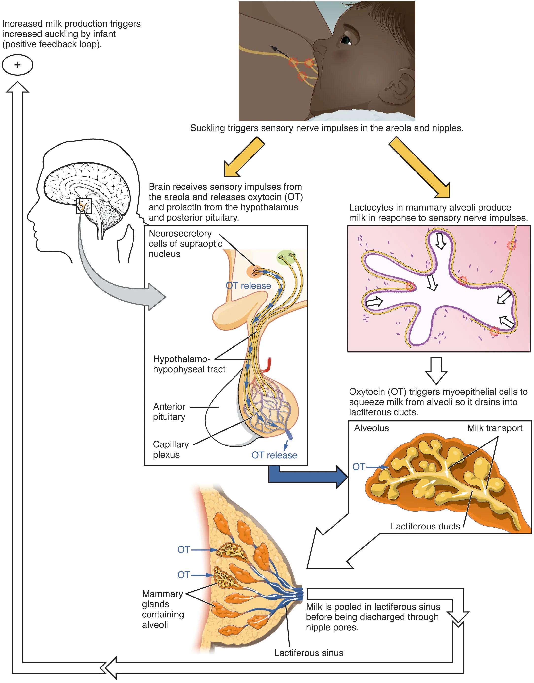
Neurosecretory cells of supraoptic nucleus: These specialized neurons, located in the hypothalamus, are responsible for synthesizing oxytocin. Upon receiving appropriate stimulation, they transmit electrical signals that culminate in the release of oxytocin from the posterior pituitary gland.
Hypothalamo-hypophysial tract: This neural pathway consists of axons originating from neurosecretory cells in the hypothalamus (including those in the supraoptic nucleus). These axons extend down into the posterior pituitary, serving as the conduit for the transport and ultimate release of neurohormones like oxytocin into the bloodstream.
Anterior pituitary: While primarily known for producing and secreting hormones such as prolactin, which is crucial for milk production, the anterior pituitary is distinct from the posterior pituitary where oxytocin is released. It receives hormonal signals from the hypothalamus via a portal system.
Capillary plexus: A network of tiny blood vessels surrounding the neurosecretory terminals in the posterior pituitary. This extensive capillary bed allows for the efficient release of hormones, such as oxytocin (OT), directly into the systemic circulation, enabling them to reach their target organs.
OT release: Refers to the secretion of oxytocin, a powerful neuropeptide and hormone, from the posterior pituitary gland. This release is triggered by nerve impulses originating from suckling, leading to a surge of oxytocin in the bloodstream.
Lactocytes in mammary alveoli produce milk in response to sensory nerve impulses: Lactocytes are the epithelial cells lining the alveoli of the mammary glands, responsible for synthesizing and secreting milk components. While prolactin primarily drives milk synthesis, the overall process is finely tuned by both hormonal and neural signals.
Oxytocin (OT) triggers myoepithelial cells to squeeze milk from alveoli so it drains into lactiferous ducts: Oxytocin acts directly on myoepithelial cells, which are specialized smooth muscle-like cells surrounding the alveoli. Their contraction forcibly expels milk from the alveoli into the intricate network of lactiferous ducts, making it available for the infant.
Alveolus: These are the small, sac-like structures within the mammary glands where milk is produced and stored. Each alveolus is lined with lactocytes and surrounded by myoepithelial cells, forming the fundamental unit of milk secretion.
Milk transport: This refers to the movement of milk from the alveoli, through the lactiferous ducts, and towards the nipple. This process is actively facilitated by the contraction of myoepithelial cells under the influence of oxytocin.
Mammary glands containing alveoli: These are the milk-producing glands located in the breast, composed of glandular tissue, fat, and connective tissue. Within the mammary glands, clusters of alveoli are organized into lobules, which drain into a system of ducts.
Lactiferous ducts: A branching network of ducts within the mammary gland that collect milk from the alveoli. These ducts converge towards the nipple, transporting milk to the lactiferous sinuses.
Lactiferous sinus: Dilated segments of the lactiferous ducts located just beneath the areola, where milk can be temporarily pooled. This pooling makes milk readily available for the infant during suckling before it is discharged through the nipple pores.
The Neuroendocrine Orchestration of Milk Ejection
The let-down reflex is an excellent example of a neuroendocrine reflex operating via a positive feedback loop. The process begins with suckling, which is a powerful sensory stimulus. Nerve impulses generated in the mechanoreceptors of the areola and nipples travel along afferent neural pathways to the spinal cord and then ascend to the hypothalamus in the brain.
Upon reaching the hypothalamus, these neural signals stimulate the neurosecretory cells, particularly those in the paraventricular and supraoptic nuclei, to synthesize and release oxytocin. Oxytocin is then transported down the hypothalamo-hypophysial tract and released from the posterior pituitary gland directly into the bloodstream.
Simultaneously, suckling also triggers the release of prolactin from the anterior pituitary, which is essential for ongoing milk production. However, it is oxytocin that is the primary hormone responsible for the milk ejection itself. Once in the systemic circulation, oxytocin travels to the mammary glands, where it specifically targets the myoepithelial cells surrounding the alveoli. The contraction of these myoepithelial cells squeezes the milk from the alveoli into the lactiferous ducts, and subsequently into the lactiferous sinuses, making it accessible to the infant.
This entire process is reinforced by a positive feedback loop: increased milk production triggers increased suckling by the infant, which in turn leads to further oxytocin and prolactin release, ensuring a continuous supply of milk as long as breastfeeding continues. This elegant physiological mechanism highlights the profound hormonal and neural adaptations that support successful lactation.
Conclusion
The intricate dance of hormones and neural signals that orchestrates the let-down reflex is fundamental to successful breastfeeding. From the initial sensory input of suckling to the precise release of oxytocin and its action on the myoepithelial cells, each step in this positive feedback loop is vital for milk ejection. Understanding these detailed neuroendocrine pathways provides invaluable insight into the physiology of lactation, equipping healthcare professionals with the knowledge to support nursing parents and address any challenges that may arise in this natural, yet complex, process.

