Delve into the intricate structural layers of the heart wall and its protective outer coverings, as detailed in this sectional view. This exploration illuminates how each distinct layer contributes to the heart’s tireless pumping function and provides essential protection. A clear understanding of these anatomical components is fundamental for comprehending cardiac physiology and various heart conditions.
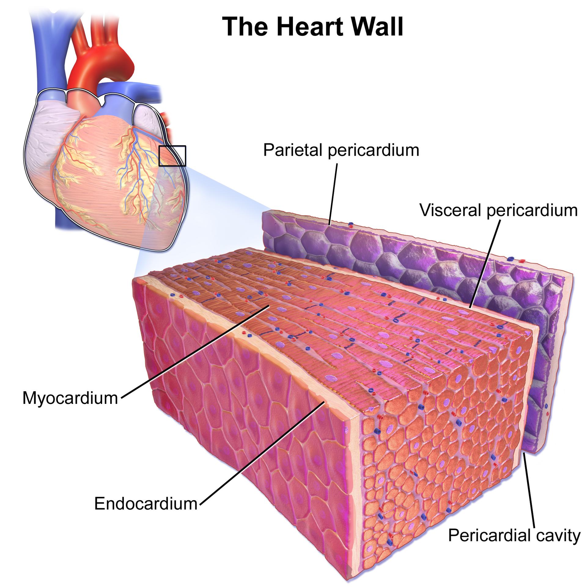
Parietal pericardium: The parietal pericardium is the tough, outer fibrous layer of the pericardium, fused to the diaphragm and great vessels, providing strong protection and anchoring the heart. It forms the outer boundary of the pericardial sac, preventing overfilling of the heart.
Visceral pericardium: Also known as the epicardium, the visceral pericardium is the innermost layer of the pericardium, directly adhering to the surface of the heart muscle. It forms the outermost layer of the heart wall itself, often containing coronary blood vessels and adipose tissue.
Myocardium: The myocardium is the thick, muscular middle layer of the heart wall, composed of specialized cardiac muscle cells. This layer is responsible for the heart’s powerful contractions, pumping blood throughout the body.
Endocardium: The endocardium is the innermost layer of the heart wall, a thin, smooth membrane that lines the heart chambers and covers the heart valves. It provides a non-thrombogenic surface for blood flow, preventing clots from forming.
Pericardial cavity: The pericardial cavity is the potential space between the parietal and visceral layers of the pericardium, containing a small amount of serous fluid. This fluid acts as a lubricant, reducing friction between the layers as the heart beats.
The heart wall, a marvel of biological engineering, is composed of three distinct layers, each with specialized functions critical to the heart’s ability to pump blood efficiently. Encasing this vital organ is the pericardium, a protective sac that anchors the heart within the chest cavity and facilitates frictionless movement. This sectional view provides an excellent opportunity to appreciate the individual contributions of these layers, from the outermost protective coverings to the innermost lining. Understanding these structural components is foundational to grasping cardiac mechanics and recognizing the impact of various diseases.
The outermost protective layer is the pericardium, which consists of a fibrous outer layer and a serous inner layer. The parietal pericardium forms the outer sac, providing robust protection and preventing the heart from over-expanding. Immediately beneath it lies the pericardial cavity, a space containing a small amount of fluid that minimizes friction as the heart beats. The inner layer of the serous pericardium, known as the visceral pericardium (or epicardium), directly adheres to the surface of the heart muscle.
Beneath the epicardium lies the formidable myocardium, the thickest and most crucial layer of the heart wall. Composed of cardiac muscle cells, the myocardium is responsible for the heart’s rhythmic contractions, generating the force needed to propel blood. The thickness of the myocardium varies across the heart’s chambers, with the left ventricle possessing the thickest wall to generate the highest pressure required for systemic circulation. Finally, the endocardium, a smooth, thin lining, covers the inner surfaces of the heart chambers and valves, preventing blood clots and ensuring laminar blood flow.
- Inflammation of the pericardium is called pericarditis, often causing chest pain.
- Myocardial infarction (heart attack) occurs when blood flow to the myocardium is blocked, leading to tissue damage.
- Infective endocarditis is a serious infection of the endocardium, often affecting the heart valves.
- The thickness of the myocardium can increase in response to conditions like high blood pressure, a condition known as hypertrophy.
A deep understanding of the heart wall and pericardium is essential for diagnosing and treating a wide range of cardiovascular conditions. For example, conditions affecting the pericardium, like pericardial effusion (excess fluid in the pericardial cavity), can impair the heart’s ability to fill with blood. Similarly, diseases targeting the myocardium, such as hypertrophic cardiomyopathy, can disrupt the heart’s pumping efficiency. The integrity and healthy function of each of these layers are paramount for overall cardiac performance and, ultimately, for life itself.
This detailed sectional view of the heart wall and pericardium provides crucial insights into the structural foundation of cardiac function. Each layer, from the protective parietal pericardium to the contractive myocardium and the smooth endocardium, plays an indispensable role in the heart’s tireless work. A comprehensive understanding of these components is vital for medical education, clinical practice, and for anyone seeking a deeper appreciation of the intricate mechanics that sustain human life.

