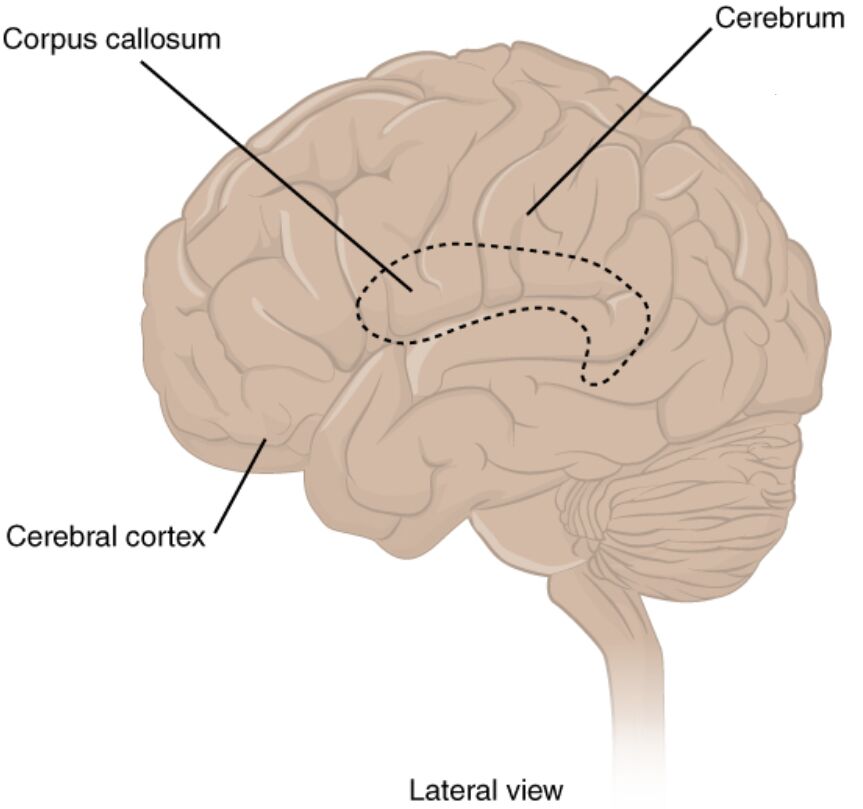
The human brain is a marvel of biological engineering, with the cerebrum serving as its largest and most prominent component. This lateral view of the cerebrum highlights key structures such as the cerebrum, corpus callosum, and cerebral cortex, offering a window into the complex workings of the central nervous system (CNS). This article provides a comprehensive exploration of these anatomical features, their functions, and their significance in maintaining bodily and cognitive health.
Cerebrum
The cerebrum is the largest part of the brain, divided into two hemispheres connected by the corpus callosum. It is responsible for higher brain functions, including thought, action, memory, and voluntary movement, making it essential for daily activities and cognitive processes.
Corpus callosum
The corpus callosum is a thick band of nerve fibers that facilitates communication between the left and right hemispheres of the cerebrum. This structure plays a critical role in integrating sensory, motor, and cognitive information, ensuring coordinated function across both sides of the brain.
Cerebral cortex
The cerebral cortex forms the outer layer of the cerebrum, characterized by its folded surface that increases its surface area. This region is pivotal for processing sensory information, initiating voluntary movements, and housing higher-order functions such as language, reasoning, and problem-solving.
Anatomical Overview of the Cerebrum
The cerebrum is a critical component of the central nervous system, responsible for orchestrating a wide range of bodily functions. Its intricate structure, visible in the lateral view, includes several key areas that work in unison to support life and cognition.
- The cerebrum occupies the upper part of the cranial cavity and is divided into four major lobes: frontal, parietal, temporal, and occipital, each with specialized roles.
- Its surface, known as the cerebral cortex, contains billions of neurons that facilitate complex neural networks essential for thought and perception.
- The folding pattern, or gyri and sulci, maximizes the cortex’s surface area within the confined space of the skull, enhancing its processing capacity.
- Blood supply to the cerebrum is maintained by the internal carotid and vertebral arteries, ensuring a constant oxygen and nutrient delivery critical for its function.
Detailed Examination of the Corpus Callosum
The corpus callosum serves as a vital bridge between the cerebral hemispheres, enabling seamless interaction. This structure is essential for maintaining brain symmetry and function.
- Comprising approximately 200 million axons, the corpus callosum transmits neural signals at high speeds, supporting interhemispheric communication.
- Damage or severance of this structure, as seen in split-brain patients, can lead to unique neurological phenomena, such as the inability to verbally name objects seen only by the left visual field.
- It plays a significant role in motor coordination, allowing both hands to perform synchronized tasks effectively.
- Developmental abnormalities, such as agenesis of the corpus callosum, can impact cognitive and motor skills, highlighting its importance.
Insights into the Cerebral Cortex
The cerebral cortex is the brain’s outer layer, critical for higher cognitive functions and sensory processing. Its complex structure supports a variety of specialized tasks.
- Composed of six layers of neurons, the cortex varies in thickness from 2 to 4 millimeters, depending on the region and its function.
- It is divided into functional areas, including the motor cortex for movement control and the sensory cortex for processing touch and pain.
- The prefrontal cortex, located in the frontal lobe, is crucial for decision-making, personality expression, and planning.
- Neuroplasticity within the cerebral cortex allows it to adapt and reorganize following injury or learning, a key aspect of brain recovery.
Clinical Relevance and Functional Importance
Understanding the anatomy of the cerebrum has significant implications for medical practice and research. The structures highlighted in this lateral view are integral to diagnosing and treating various neurological conditions.
- The cerebrum’s role in cognition makes it a focus for studying disorders like Alzheimer’s disease, where cortical atrophy is a common feature.
- The corpus callosum’s integrity is assessed in conditions such as multiple sclerosis, where demyelination can disrupt interhemispheric communication.
- The cerebral cortex’s sensory and motor areas are evaluated in stroke patients to determine the extent of impairment and guide rehabilitation.
- Advanced imaging techniques, such as MRI, provide detailed views of these structures, aiding in precise surgical planning for brain tumors or epilepsy.
Conclusion
The lateral view of the cerebrum offers a fascinating glimpse into the brain’s architecture, with the cerebrum, corpus callosum, and cerebral cortex playing pivotal roles in human function. These structures not only support essential physiological processes but also underpin our capacity for thought, movement, and interaction with the world. Exploring their anatomy deepens our appreciation of the brain’s complexity and informs ongoing efforts in medical science to enhance health and treat neurological disorders effectively.

