The human body is a complex and fascinating network of organ systems, each playing a vital role in maintaining health and functionality. This detailed medical image provides an insightful overview of the Lymphatic System, Respiratory System, Digestive System, Urinary System, Male Reproductive System, and Female Reproductive System, highlighting their key components and functions. Whether you’re exploring anatomy for educational purposes or personal interest, this guide offers a comprehensive look at how these systems work together to sustain life.
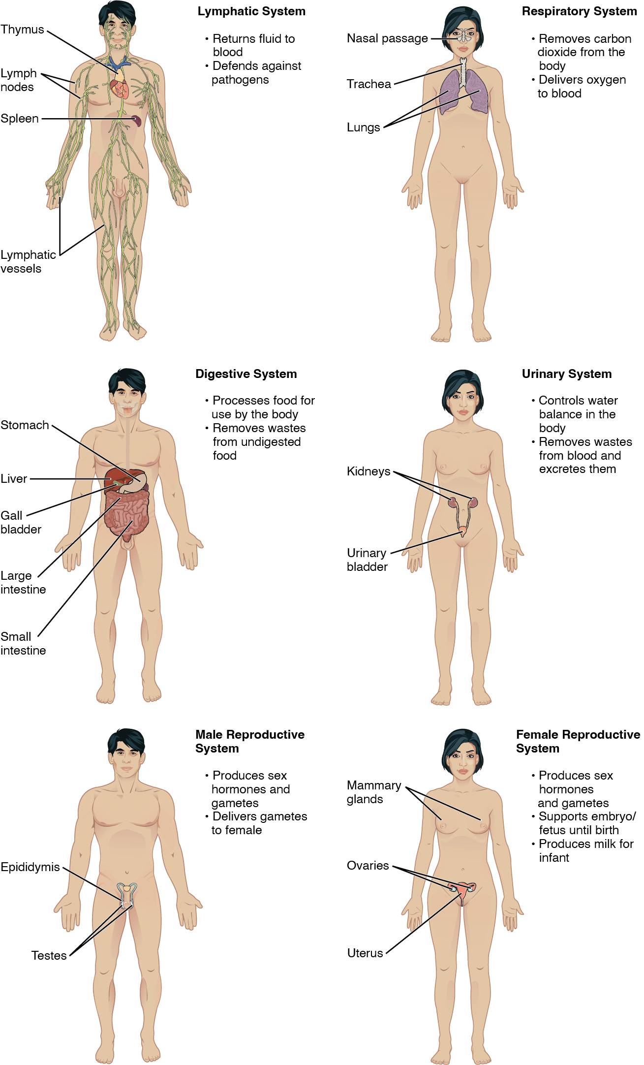
- Thymus: The thymus is a small organ located in the upper chest, crucial for the development of T-lymphocytes that help fight infections. It plays a significant role in the immune system, particularly during childhood and adolescence.
- Lymph nodes: These small, bean-shaped structures are scattered throughout the body and filter lymph fluid, trapping bacteria, viruses, and other pathogens. They are essential for activating the immune response to protect against disease.
- Spleen: Positioned in the upper left abdomen, the spleen filters blood, removes old or damaged red blood cells, and stores white blood cells. It also aids in immune function by producing antibodies.
- Lymphatic vessels: These extensive networks transport lymph fluid back into the bloodstream, helping maintain fluid balance and supporting immune responses. They connect various parts of the body, ensuring efficient waste removal.
- Nasal passage: The nasal passage serves as the entry point for air, filtering, warming, and humidifying it before it reaches the lungs. It also plays a role in the sense of smell, enhancing respiratory efficiency.
- Trachea: Known as the windpipe, the trachea is a tube that carries air from the larynx to the bronchi, lined with cartilage rings to keep it open. It protects the airway and prevents collapse during breathing.
- Lungs: The lungs are large, spongy organs responsible for oxygenating blood and expelling carbon dioxide through respiration. They contain millions of alveoli where gas exchange occurs.
- Stomach: The stomach is a muscular organ that breaks down food with gastric juices, including hydrochloric acid and enzymes. It temporarily stores food and initiates protein digestion.
- Liver: The liver, located in the upper right abdomen, detoxifies blood, produces bile for fat digestion, and stores nutrients like glycogen. It also synthesizes proteins essential for blood clotting.
- Gall bladder: This small organ stores and concentrates bile produced by the liver, releasing it into the small intestine to aid fat digestion. It plays a key role in the emulsification of dietary fats.
- Large intestine: The large intestine absorbs water and electrolytes from undigested food, forming feces for elimination. It houses beneficial bacteria that aid in vitamin production.
- Small intestine: This long, coiled organ is where most nutrient absorption occurs, aided by enzymes and a vast surface area. It completes the digestion process started in the stomach.
- Kidneys: The kidneys filter blood to remove waste and excess water, forming urine, and regulate electrolyte balance. They are vital for maintaining blood pressure and red blood cell production.
- Urinary bladder: The urinary bladder stores urine produced by the kidneys until it is excreted through the urethra. Its muscular walls allow it to expand and contract as needed.
- Testes: The testes produce sperm and male sex hormones like testosterone, which regulate secondary sexual characteristics. They are housed in the scrotum to maintain optimal temperature for sperm production.
- Epididymis: The epididymis is a coiled tube where sperm mature and are stored before ejaculation. It also aids in the transport of sperm during reproduction.
- Mammary glands: These glands in the female breast produce milk to nourish infants after childbirth. They are influenced by hormones like prolactin and oxytocin.
- Ovaries: The ovaries produce eggs and female sex hormones such as estrogen and progesterone, regulating the menstrual cycle. They are essential for reproduction and hormonal balance.
- Uterus: The uterus supports the development of a fetus during pregnancy, with thick muscular walls for labor. It sheds its lining monthly in the absence of pregnancy.
Lymphatic System: The Body’s Defense Network
This system is crucial for maintaining fluid balance and defending against pathogens. The thymus releases hormones like T3 and T4 to support immune cell maturation, while lymph nodes act as checkpoints, filtering lymph and initiating immune responses. The spleen removes old blood cells and produces antibodies, enhancing overall immunity. Lymphatic vessels ensure continuous circulation of lymph, preventing fluid buildup and supporting the body’s defense mechanism.
Respiratory System: Oxygen and Carbon Dioxide Exchange
The respiratory system ensures the body receives oxygen and expels carbon dioxide. The nasal passage filters air and protects the lungs from debris, while the trachea provides a clear airway with its cartilaginous structure. The lungs facilitate gas exchange, with alveoli optimizing oxygen uptake and carbon dioxide removal, critical for cellular respiration.
Digestive System: Processing Nutrients and Waste
This system breaks down food for energy and eliminates waste. The stomach uses hydrochloric acid to digest proteins, while the liver detoxifies blood and produces bile. The gall bladder releases bile to emulsify fats, and the large intestine absorbs water, housing gut microbiota. The small intestine maximizes nutrient absorption with its extensive surface area.
Urinary System: Maintaining Fluid and Waste Balance
The urinary system regulates water and removes waste from the blood. The kidneys filter approximately 120-150 quarts of blood daily, producing urine to excrete waste. The urinary bladder stores this urine, releasing it through a controlled process, ensuring homeostasis.
Male Reproductive System: Hormones and Gamete Production
This system is responsible for producing male gametes and hormones. The testes synthesize testosterone and sperm, while the epididymis matures and stores sperm for delivery. These components work together to support reproduction and male secondary characteristics.
Female Reproductive System: Supporting Reproduction and Lactation
The female system supports pregnancy and lactation. The mammary glands produce milk under hormonal control, while the ovaries release eggs and hormones like estrogen. The uterus provides a nurturing environment for fetal development, completing the reproductive cycle.
Understanding these organ systems offers a foundation for appreciating human physiology. Each system’s unique functions contribute to a harmonious balance, ensuring survival and health. Exploring their interactions can deepen your knowledge of the body’s remarkable capabilities.

