The disposable device shown in the image is an electrocardiogram (EKG or ECG) electrode, a fundamental component in modern cardiology used to detect the heart’s electrical activity. These sensors act as transducers, converting the ionic currents generated by the heart muscle into electron currents that can be interpreted by a monitoring machine. By adhering securely to the patient’s skin, these electrodes ensure the transmission of clear, high-fidelity signals, which are essential for diagnosing heart conditions ranging from minor irregular heartbeats to life-threatening cardiac events.
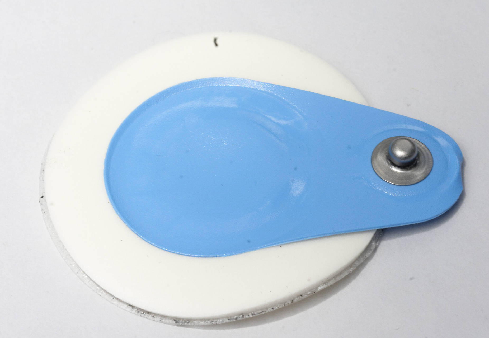
Anatomical Components of the EKG Sensor
Metal Snap Connector:
The prominent silver stud located on the blue tab is the connection interface. It allows the lead wires from the EKG machine to snap securely onto the electrode, ensuring a stable pathway for electrical signals to travel from the sensor to the monitor.
Adhesive Foam Backing:
The white, circular outer ring is constructed from a medical-grade foam material coated with a hypoallergenic adhesive. This component anchors the electrode to the patient’s skin, isolating the sensor from external movement and preventing “motion artifacts” that could distort the cardiac reading.
Conductive Gel Matrix:
Although hidden beneath the center of the snap, this is the functional core of the device, typically containing a silver/silver chloride (Ag/AgCl) electrolyte gel. This wet or solid gel reduces the natural electrical resistance (impedance) of the skin, allowing the faint electrical impulses of the heart to be detected accurately.
Physiology of Cardiac Electrical Detection
To understand how this simple sensor functions, one must first understand the physiology of the heart. The heart is not just a mechanical pump; it is driven by a complex electrical system. The heartbeat originates in the sinoatrial (SA) node, the heart’s natural pacemaker, which generates an electrical impulse. This impulse creates a wave of depolarization that spreads through the atria and ventricles, causing the muscle fibers to contract and pump blood. Because the human body is a volume conductor, these electrical currents radiate from the heart to the surface of the skin.
However, dry skin is a poor conductor of electricity. This is where the EKG electrode plays a crucial role. The silver/silver chloride chemistry within the sensor is designed to bridge the gap between the biological ionic current in the body and the electronic current required by the medical equipment. When the electrode is placed on the skin, the conductive gel penetrates the dead outer layer of the epidermis (stratum corneum), significantly lowering skin impedance. This allows the millivolt-level signals generated by the heart to be picked up clearly without excessive noise or interference.
The specific placement of these sensors is vital for a standard 12-lead EKG. Ten electrodes are typically placed on the limbs and across the chest (precordium). By measuring the voltage difference between these various points, the EKG machine creates a three-dimensional representation of the heart’s electrical activity. This data is critical for identifying pathology. For instance, if the electrical signal is blocked or delayed, it may indicate a heart block. If the electrical activity is chaotic, it suggests fibrillation.
- Key features of a high-quality EKG sensor include:
- Ag/AgCl Composition: Provides the most stable baseline and lowest noise for accurate signal tracking.
- Aggressive Adhesion: Ensures the sensor does not peel off during stress testing or diaphoretic (sweaty) conditions.
- Hypoallergenic Materials: Reduces the risk of contact dermatitis during long-term monitoring.
- Radiolucency: Some sensors are designed to be invisible to X-rays, allowing for imaging without removing the leads.
Clinical Significance: Detecting Arrhythmias and Ischemia
The primary clinical application of this sensor is the diagnosis of cardiovascular diseases, specifically arrhythmia and myocardial ischemia. An arrhythmia refers to an abnormal heart rhythm, where the heart may beat too fast (tachycardia), too slow (bradycardia), or irregularly. For example, in Atrial Fibrillation (AFib), the upper chambers of the heart quiver chaotically rather than contracting normally. The EKG electrode picks up these disorganized electrical signals, which appear as irregular, rapid squiggles between the heartbeats on the monitor. Without the high-fidelity transmission provided by the electrode, these subtle changes in the waveform might be missed, leading to a misdiagnosis.
Furthermore, these sensors are indispensable in the acute setting of a heart attack, or myocardial infarction. When blood flow to a part of the heart is blocked, the muscle begins to die, changing how it conducts electricity. On an EKG tracing, this often manifests as an elevation or depression of the “ST segment.” The electrode must maintain a stable baseline to allow physicians to measure these deviations accurately. Even a millimeter of shift on the graph paper can determine whether a patient needs immediate catheterization or medication management. Therefore, the quality of the sensor and the skin preparation before application are as important as the sophistication of the monitoring machine itself.
Conclusion
While it may appear to be a simple disposable item, the EKG electrode is a sophisticated piece of medical technology that serves as the gateway to cardiac diagnostics. Its design—integrating a conductive electrolyte with a stable adhesive platform—overcomes the body’s natural resistance to reveal the internal electrical workings of the heart. From routine check-ups to emergency room resuscitation, the reliability of the data displayed on a cardiac monitor depends entirely on the integrity of this small interface, making it a cornerstone of cardiovascular care.

