The human body is a complex and fascinating structure, with muscles playing a critical role in movement and stability. This article delves into the anatomical details of the pectoralis major, deltoid, and latissimus dorsi muscles, as depicted in the provided medical image. These muscles are essential for upper body strength and mobility, making them a key focus for those studying human anatomy or seeking to understand musculoskeletal health. By exploring their locations, functions, and interconnections, readers can gain a deeper appreciation of how these muscles contribute to everyday activities and physical fitness.
Introduction to the Labeled Muscles
This section provides an overview of the key muscles highlighted in the image, offering a foundation for understanding their roles. The following details break down each labeled muscle to enhance your anatomical knowledge.
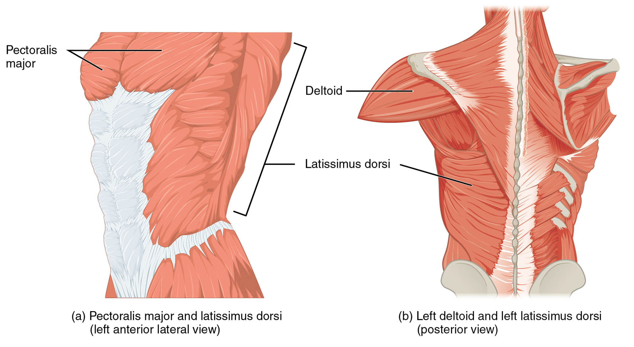
- Pectoralis major: This large chest muscle is located on the anterior lateral view of the torso, covering the upper rib cage. It is primarily responsible for flexing, adducting, and internally rotating the humerus, playing a vital role in arm movements.
- Deltoid: Positioned over the shoulder joint in the posterior view, this muscle forms the rounded contour of the shoulder. It enables abduction, flexion, and extension of the arm, providing essential support for shoulder stability.
- Latissimus dorsi: Found extending across the lower and middle back in the posterior view, this broad muscle aids in extending, adducting, and internally rotating the arm. It is crucial for powerful pulling motions and upper body strength.
Detailed Anatomical Overview
This part expands on the structural and functional aspects of the muscles, offering a comprehensive look at their anatomy. The pectoralis major, deltoid, and latissimus dorsi are integral to the upper body’s musculoskeletal framework, each with unique characteristics.
- The pectoralis major originates from the clavicle, sternum, and upper ribs, inserting into the humerus. Its fan-shaped structure allows for a wide range of shoulder movements, supported by its innervation from the medial and lateral pectoral nerves.
- The deltoid is a thick, triangular muscle with origins at the clavicle, acromion, and spine of the scapula, inserting into the deltoid tuberosity. Its three distinct heads (anterior, middle, and posterior) work synergistically to provide multidirectional arm motion.
- The latissimus dorsi originates from the thoracolumbar fascia, iliac crest, and lower thoracic vertebrae, converging to insert into the intertubercular groove of the humerus. Innervated by the thoracodorsal nerve, it contributes significantly to back and arm strength.
Functional Roles in the Body
This section highlights the practical applications of these muscles, emphasizing their importance in physical activities. The pectoralis major, deltoid, and latissimus dorsi are not only anatomical structures but also key players in human movement.
- The pectoralis major is heavily engaged in pushing actions, such as bench presses, and assists in breathing by stabilizing the chest wall. Its robust blood supply from the pectoral branches of the thoracoacromial artery ensures efficient function.
- The deltoid supports overhead lifting and throwing motions, with its middle fibers particularly active during lateral raises. This muscle’s rich vascularization aids in rapid recovery after intense use.
- The latissimus dorsi powers swimming, rowing, and climbing by facilitating arm extension and adduction. Its extensive connection to the spine enhances posture and core stability during these activities.
Clinical Significance and Considerations
This part addresses the relevance of these muscles in a medical context, providing insights into their health implications. Understanding the pectoralis major, deltoid, and latissimus dorsi can aid in diagnosing and treating related conditions.
- Injuries to the pectoralis major, such as tears from weightlifting, can lead to pain and reduced arm function, often requiring surgical intervention. Rehabilitation focuses on restoring range of motion and strength.
- The deltoid may suffer from strains or rotator cuff-related issues, impacting shoulder mobility. Physical therapy often targets this muscle to alleviate pain and prevent chronic damage.
- The latissimus dorsi can be affected by overuse injuries in athletes, leading to back pain or limited arm movement. Stretching and strengthening exercises are commonly recommended for recovery.
As we conclude this exploration, the pectoralis major, deltoid, and latissimus dorsi stand out as vital components of the upper body’s muscular system. Their intricate design and coordinated functions enable a wide range of motions, from lifting to pulling, while also supporting overall posture and stability. Whether for academic study or personal health, understanding these muscles provides valuable insights into human anatomy and its practical applications. For further learning, consider examining their nerve supply and vascular connections in greater detail to appreciate their full complexity.

