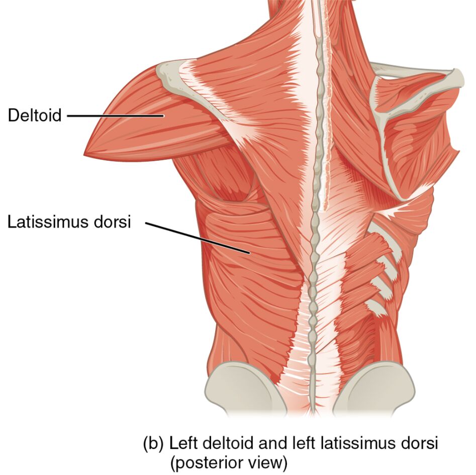
The human body is a remarkable structure, with muscles playing a crucial role in movement and stability. This article delves into the anatomy of the left deltoid and latissimus dorsi muscles, as depicted in a posterior view image. These muscles are essential for upper body mobility, and understanding their functions can enhance knowledge of musculoskeletal health. The image provides a clear illustration of their locations and relationships, offering valuable insights into their roles in shoulder and back movements.
Introduction to Labeled Muscles
This section explores the key muscles highlighted in the image, providing a detailed look at their anatomical significance. Each labeled muscle is critical to the mechanics of the upper body, and their functions are worth examining closely.
- Deltoid:
The deltoid is a large, triangular muscle covering the shoulder joint, originating from the clavicle and scapula.
It is responsible for arm abduction, flexion, and extension, making it vital for a wide range of upper limb movements. - Latissimus dorsi:
The latissimus dorsi is a broad, flat muscle spanning the middle and lower back, originating from the spine and iliac crest.
It facilitates arm adduction, extension, and internal rotation, playing a key role in pulling and lifting actions.
This part dives deeper into the structure and function of the muscles shown in the image. It offers a comprehensive overview to aid in understanding their contributions to body mechanics.
The deltoid muscle, located at the shoulder, is composed of three distinct heads—anterior, middle, and posterior—that work together to provide a full range of motion. Originating from the superior surfaces of the scapula and clavicle, it inserts into the deltoid tuberosity of the humerus, enabling superior movement of the arm. Its strength and versatility make it a cornerstone of shoulder stability and mobility.
The latissimus dorsi, in contrast, extends across the lower and middle back, with origins from the thoracolumbar fascia, iliac crest, and lower thoracic vertebrae. Inserting into the intertubercular groove of the humerus, it drives inferior movements of the arm, such as pulling the arm downward or backward. Together, these muscles exemplify the intricate balance of the musculoskeletal system, supporting both dynamic and static postures.
Their coordination is essential for activities like lifting, throwing, or swimming, where the deltoid initiates upward motion and the latissimus dorsi counters with downward force. This synergy highlights the importance of maintaining muscle health through proper exercise and care, as imbalances can lead to strain or injury.
Functional Roles in Movement
This section examines how these muscles contribute to specific movements. It provides insight into their practical applications in everyday activities.
The deltoid’s primary function is to move the humerus superiorly, a motion critical for raising the arm overhead or laterally.
Its origin from the scapula and clavicle ensures a stable base for these actions, supported by its robust muscular structure.
The latissimus dorsi, originating from the middle and lower back, moves the humerus inferiorly, essential for pulling motions.
This muscle’s extensive coverage allows it to generate significant power, aiding in activities like rowing or climbing.
Both muscles work in opposition to maintain balance and control, with the deltoid elevating the arm and the latissimus dorsi providing a counterforce. This dynamic interplay is crucial for athletes and individuals engaging in physical labor, underscoring the need for targeted strengthening exercises.
Clinical Relevance and Muscle Health
This part addresses the practical implications of understanding these muscles. It offers guidance on maintaining their health and recognizing potential issues.
Healthy deltoid and latissimus dorsi muscles are vital for preventing injuries such as rotator cuff tears or lower back strain.
Regular stretching and strengthening exercises can enhance their resilience, particularly for those with active lifestyles.
Injury to the deltoid may result from overuse, leading to inflammation or tears that limit shoulder mobility.
Similarly, latissimus dorsi strain, often from repetitive pulling, can cause pain and restrict back movement, necessitating rest and rehabilitation.
Proper warm-ups and ergonomic practices can mitigate these risks, ensuring long-term muscle function. Consulting a healthcare professional is advisable for persistent discomfort, as early intervention can prevent chronic conditions.
Conclusion
The left deltoid and latissimus dorsi muscles, as illustrated in the posterior view, are integral to the upper body’s strength and mobility. This article has explored their anatomical locations, functional roles, and clinical significance, providing a solid foundation for understanding their contributions. Maintaining these muscles through balanced exercise and mindful practices is key to supporting overall physical health. By appreciating their intricate design and interplay, one can better appreciate the complexity of human movement and the importance of musculoskeletal care.

