The tonsils are an integral part of the immune system, strategically located in the throat to protect against inhaled or ingested pathogens. These lymphoid tissues, including the palatine, pharyngeal, and lingual tonsils, act as the first line of defense by trapping bacteria and viruses, initiating immune responses to maintain respiratory and digestive health. This detailed anatomical illustration provides a clear view of their positions and surrounding structures, offering valuable insights into their protective roles.
Labeled Components of the Tonsils
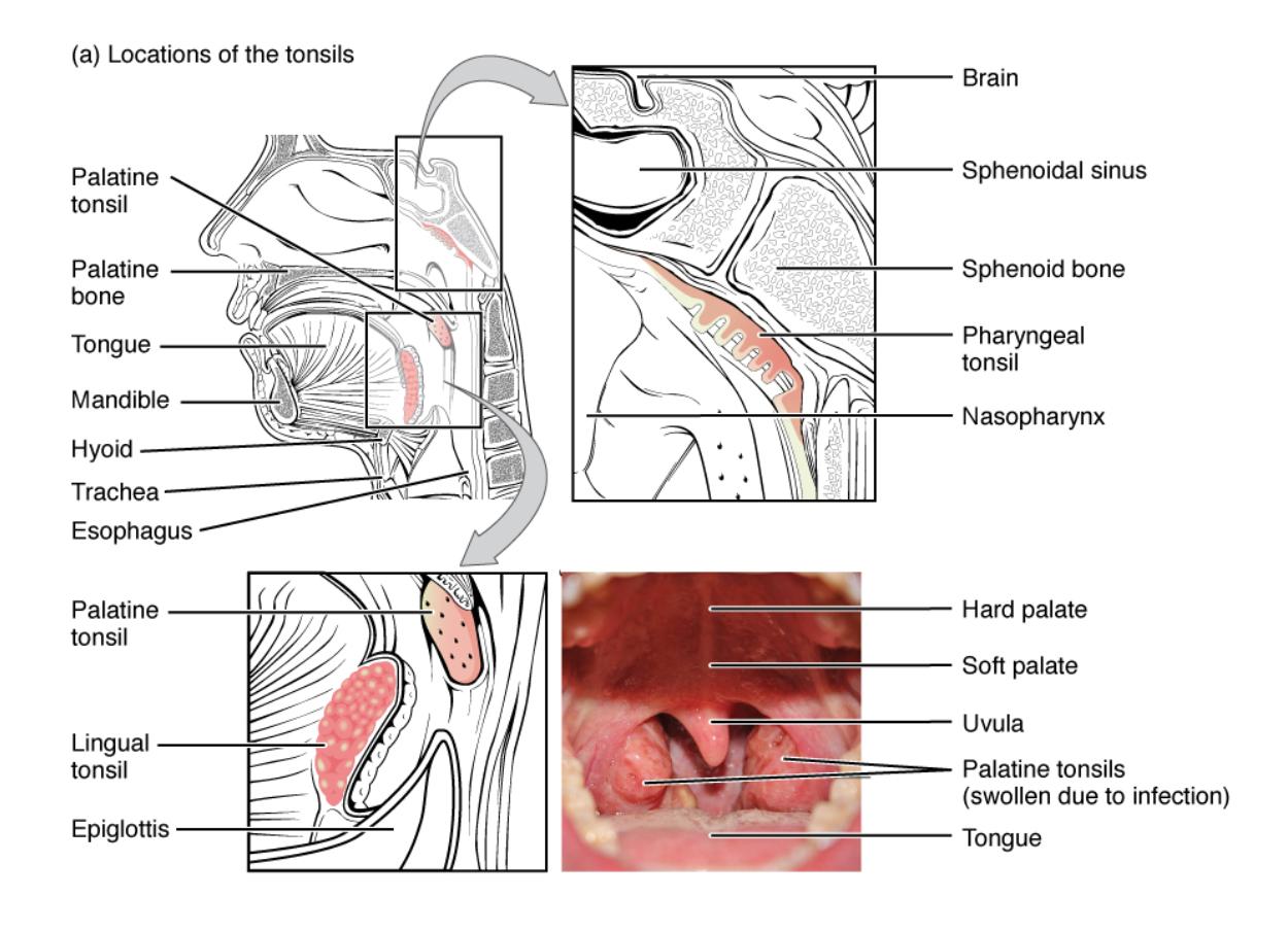
Palatine tonsil: These tonsils are situated on either side of the pharynx, at the back of the throat, and are commonly visible when the mouth is open. They play a key role in trapping pathogens entering through the mouth, contributing to local immunity by producing antibodies.
Palatine bone: Forming part of the hard palate and nasal cavity, this bone provides structural support to the palatine tonsils. It serves as an anchor, stabilizing the tonsillar tissue within the oral cavity.
Tongue: Located at the base of the mouth, the tongue aids in swallowing and speech while being near the lingual tonsils. Its proximity allows it to assist in immune surveillance by interacting with tonsillar lymphoid tissue.
Mandible: This lower jawbone frames the oral cavity, supporting the tongue and lingual tonsils. It provides a protective boundary, ensuring the tonsils remain in their functional position.
Hyoid: A U-shaped bone below the mandible, the hyoid supports the tongue and larynx, indirectly stabilizing the lingual tonsils. It plays a role in maintaining the structural integrity of the throat region.
Trachea: This airway tube extends from the larynx to the bronchi, positioned below the tonsillar region. It is protected by the tonsils’ immune activity against inhaled pathogens.
Esophagus: Located behind the trachea, this tube transports food to the stomach and is guarded by the tonsils against ingested microbes. Its proximity enhances the tonsils’ protective function.
Lingual tonsil: Found at the base of the tongue, these tonsils are less visible but crucial for immune defense in the oral cavity. They help neutralize pathogens entering through the mouth, complementing the palatine tonsils.
Epiglottis: This cartilage flap covers the trachea during swallowing, preventing food entry into the airway. Its position near the lingual tonsils aids in coordinating immune and swallowing functions.
Brain: Positioned above the nasal cavity, the brain is indirectly protected by the pharyngeal tonsil’s role in filtering nasal pathogens. It highlights the tonsils’ broader protective scope within the head.
Sphenoidal sinus: Located in the sphenoid bone, this air-filled cavity near the pharyngeal tonsil drains mucus and traps pathogens. It supports the tonsil’s function by filtering air before it reaches deeper structures.
Sphenoid bone: This bone forms part of the skull base, housing the sphenoidal sinus and supporting the pharyngeal tonsil. It provides a stable foundation for the upper tonsillar region.
Pharyngeal tonsil: Situated on the roof of the nasopharynx, this tonsil, also known as the adenoid, filters inhaled air. It is critical for immune defense in the nasal passages, especially in children.
Nasopharynx: The upper part of the pharynx behind the nasal cavity, it hosts the pharyngeal tonsil. This area is a key entry point for air, making the tonsil’s location strategic for pathogen capture.
Hard palate: Forming the anterior roof of the mouth, the hard palate supports the oral cavity’s structure near the palatine tonsils. It aids in separating the oral and nasal cavities, enhancing tonsillar function.
Soft palate: This flexible posterior part of the palate, near the uvula, assists in closing off the nasopharynx during swallowing. It works with the palatine tonsils to protect against ingested pathogens.
Uvula: The small projection hanging from the soft palate, the uvula helps prevent food from entering the nasopharynx. Its proximity to the palatine tonsils supports their immune role during swallowing.
Palatine tonsils (swollen due to infection): These tonsils, shown enlarged in the image, indicate inflammation often caused by bacterial or viral infections like tonsillitis. Swelling can obstruct breathing and swallowing, necessitating medical attention.
Tongue: Reiterated in the oral view, it underscores its role near the lingual and palatine tonsils. Its movement aids in clearing pathogens trapped by the tonsils.
Anatomical Overview of the Tonsils
The tonsils form a protective ring around the throat, each with a specific location and function.
- The palatine tonsils flank the pharynx, easily visible and prone to swelling during infections.
- The pharyngeal tonsil, or adenoid, guards the nasopharynx, filtering air from the nasal passages.
- Lingual tonsils at the tongue base complement oral immunity, working with the epiglottis.
- The hard and soft palates provide structural support, separating oral and nasal cavities.
- The uvula and tongue assist in swallowing, coordinating with tonsillar defenses.
- The sphenoidal sinus and bone protect the upper airway, supporting the pharyngeal tonsil.
This illustration highlights their strategic placement for immune surveillance.
Physiological Roles of the Tonsils
The tonsils contribute significantly to the body’s first line of defense against pathogens.
- Palatine tonsils trap ingested microbes, producing antibodies like IgA to neutralize threats.
- The pharyngeal tonsil filters inhaled air, reducing the risk of respiratory infections.
- Lingual tonsils enhance oral immunity, working with saliva to clear pathogens.
- The epiglottis and uvula coordinate swallowing, preventing pathogen entry into the airway.
- The sphenoidal sinus drains mucus, aiding the pharyngeal tonsil in air filtration.
- Swollen palatine tonsils signal infection, prompting immune activation or medical intervention.
This collaborative effort ensures comprehensive throat protection.
Clinical Significance of Tonsil Anatomy
Tonsil anatomy is crucial for diagnosing and treating related conditions.
- Swollen palatine tonsils may indicate tonsillitis, often requiring antibiotics or removal.
- Pharyngeal tonsil enlargement, or adenoid hypertrophy, can obstruct breathing in children.
- Lingual tonsil inflammation may cause discomfort, affecting swallowing and speech.
- The hard and soft palates’ support can be assessed in cases of tonsillar displacement.
- The uvula’s position is key in evaluating airway obstruction from tonsil swelling.
- The sphenoidal sinus’s health impacts the pharyngeal tonsil’s effectiveness against sinusitis.
This knowledge guides clinical management of tonsil-related issues.
Developmental and Immune Functions
Tonsils develop early, adapting to immune needs throughout life.
- Palatine tonsils peak in childhood, declining in size with age but retaining function.
- The pharyngeal tonsil is most active in young children, shrinking after puberty.
- Lingual tonsils mature with the tongue, supporting lifelong oral immunity.
- The epiglottis and uvula refine swallowing coordination as the tonsils develop.
- The sphenoidal sinus and bone grow to enhance upper airway protection.
- Swollen tonsils reflect an active immune response, common in early infections.
This adaptability ensures ongoing immune support.
The tonsils’ strategic locations, as depicted in this anatomical view, underscore their vital role in immune defense. Whether filtering air or food-borne pathogens, their collaboration with surrounding structures like the palate and uvula highlights their importance, making them a key focus for understanding throat health and managing infections effectively.

