Understanding the complex network of arteries in the human leg is essential for recognizing how oxygen-rich blood reaches the lower extremities to support mobility and tissue health. This anatomical guide explores the major vascular pathways, from the groin to the foot, highlighting the critical roles each vessel plays in the peripheral circulatory system.
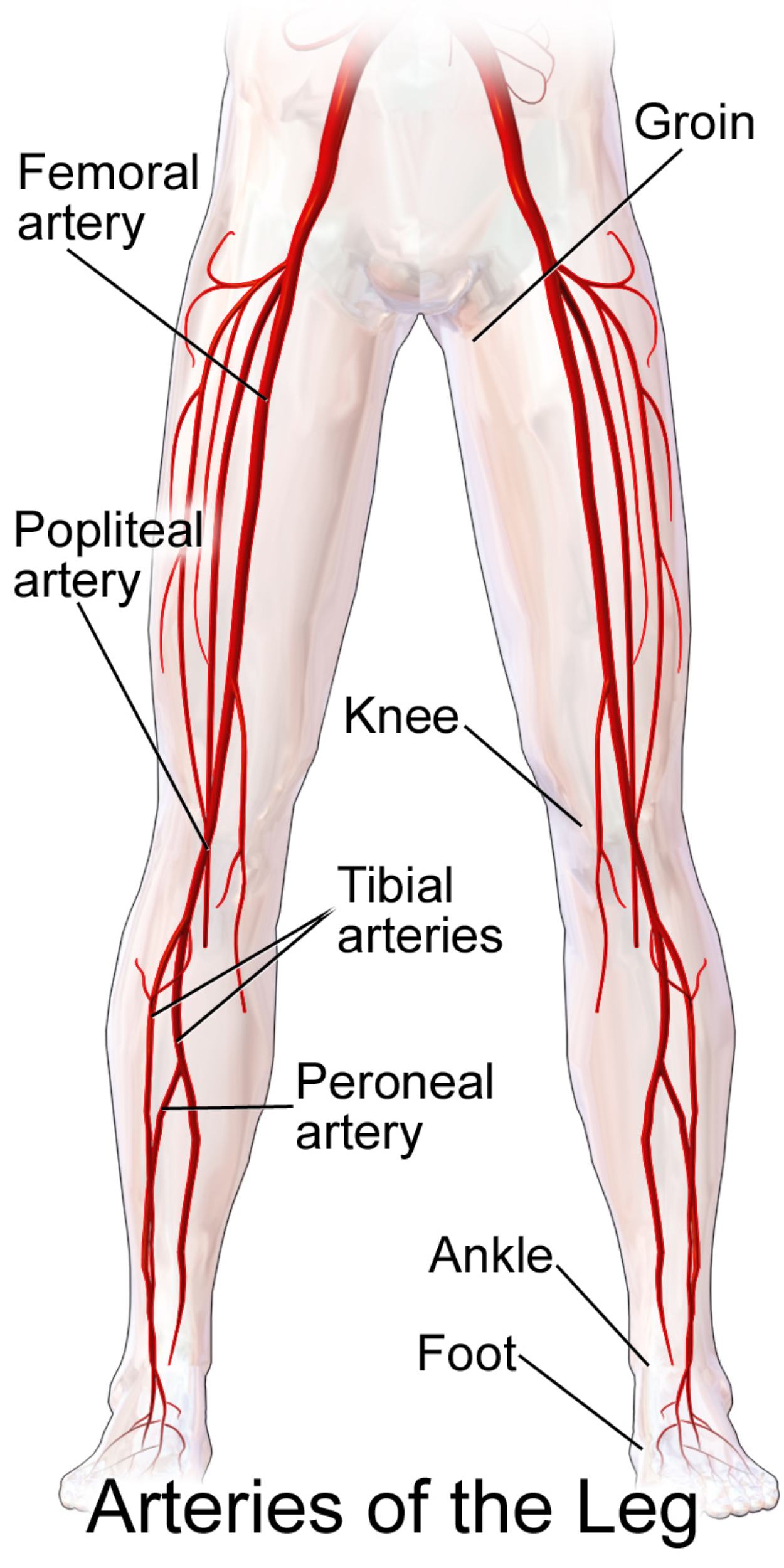
Groin: The groin marks the transition point where the external iliac artery becomes the common femoral artery as it passes under the inguinal ligament. This region is a common site for clinical procedures, such as arterial catheterization or pulse palpation, due to the vessel’s proximity to the surface.
Femoral artery: The femoral artery is the primary blood vessel supplying the thigh and is a direct continuation of the common femoral artery. It travels deep within the thigh muscles, eventually passing through the adductor canal to reach the posterior aspect of the leg.
Popliteal artery: Located behind the knee joint within the popliteal fossa, this artery is the continuation of the femoral artery. It is responsible for supplying blood to the knee joint and the muscles of the calf through various smaller branches.
Knee: The knee serves as a major anatomical landmark where the popliteal artery transitions into the smaller vessels of the lower leg. Significant collateral circulation exists around this joint to ensure blood flow remains constant even during deep flexion or movement.
Tibial arteries: These vessels consist of the anterior and posterior tibial arteries, which branch off from the popliteal artery below the knee. They are vital for supplying the anterior and posterior compartments of the lower leg, providing the necessary pressure to reach the distal extremities.
Peroneal artery: Also known as the fibular artery, this vessel branches from the posterior tibial artery and runs along the medial side of the fibula. It provides essential nourishment to the lateral compartment of the leg and the muscles surrounding the ankle.
Ankle: The ankle region acts as a terminal junction where the tibial and peroneal arteries converge and transition into the foot’s vascular network. Clinicians often check the posterior tibial pulse at the medial malleolus of the ankle to assess overall vascular health in the limb.
Foot: The arteries of the foot, including the dorsalis pedis and plantar arteries, represent the most distal part of the leg’s arterial system. These vessels form complex arches that ensure high-pressure blood reaches the toes and the soft tissues of the sole.
The Physiological Pathway of Blood Flow
The journey of blood to the lower limbs begins at the abdominal aorta, which bifurcates into the common iliac arteries before narrowing into the vessels shown in the diagram. This high-pressure system is designed to deliver oxygenated blood to demanding muscle groups, such as the quadriceps and gastrocnemius, which are essential for walking and running. The structural integrity of these vessel walls, composed of endothelial cells and smooth muscle, allows for the regulation of blood pressure and flow based on the body’s physical activity levels.
Clinical conditions often arise when these pathways become obstructed, a condition known as peripheral artery disease (PAD). This typically occurs due to atherosclerosis, where fatty deposits or plaques build up inside the arterial walls, narrowing the lumen and restricting flow. If left untreated, reduced circulation can lead to symptoms such as intermittent claudication—pain that occurs during exercise—or in severe cases, critical limb ischemia, which threatens the viability of the leg.
Maintaining the health of these arteries involves a combination of lifestyle choices and medical management. Because the legs are furthest from the heart, they are often the first to show signs of systemic vascular issues. Several factors contribute to the long-term viability of these vessels:
- Regular cardiovascular exercise to promote collateral circulation.
- Strict management of blood glucose levels, particularly in diabetic patients.
- Cessation of smoking, which is a primary risk factor for arterial wall damage.
- Monitoring cholesterol levels and blood pressure to prevent plaque buildup.
Advanced diagnostic imaging, such as Doppler ultrasound or CT angiography, allows medical professionals to visualize these arteries in real-time. By mapping the blood flow through the femoral and tibial systems, doctors can identify specific blockages and determine whether medical management or surgical intervention, such as stenting or bypass grafting, is necessary. Proactive care ensures that the peripheral tissues remain healthy and functional throughout a person’s life.
The intricate network of the leg’s arterial system is a marvel of biological engineering, ensuring that even the furthest tissues of the toes receive the nutrients necessary for survival. By understanding the anatomy from the groin to the foot, individuals and healthcare providers can better appreciate the signs of vascular health and take proactive steps to prevent chronic diseases. A healthy arterial system is the foundation of mobility, allowing the body to maintain an active and independent lifestyle.

