The image provided offers a detailed cross-sectional view of normal lung tissue, showcasing its intricate layers and components. This anatomical structure is essential for understanding the respiratory system’s functionality, providing a foundation for studying both healthy and diseased states. Below, the labeled parts of the image are explained to give a comprehensive insight into the tissue’s composition.
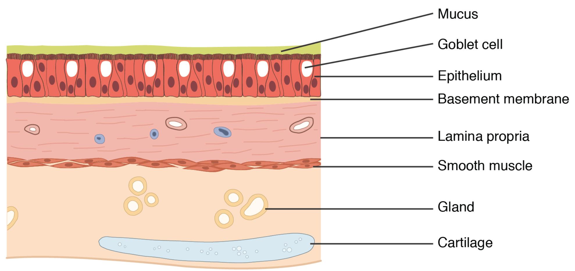
- Mucus: This sticky substance coats the surface of the lung tissue, trapping dust and pathogens to protect the respiratory tract. It is continuously produced and moved by cilia to maintain a clean airway.
- Goblet cell: These specialized cells within the epithelium secrete mucus, playing a critical role in maintaining the airway’s moisture and defense. Their presence ensures a constant supply of mucus to protect underlying tissues.
- Epithelium: This outermost layer consists of tightly packed cells that form a protective barrier, facilitating gas exchange and preventing external invaders. It contains ciliated cells that help move mucus and debris out of the lungs.
- Basement membrane: This thin layer anchors the epithelium to the underlying connective tissue, providing structural support and stability. It also regulates the exchange of substances between the epithelium and lamina propria.
- Lamina propria: Composed of loose connective tissue, this layer contains blood vessels and nerves, supporting the epithelium and aiding in nutrient supply. It also houses immune cells to respond to local infections.
- Smooth muscle: These involuntary muscles regulate airway diameter, allowing the lungs to adjust airflow during breathing. Contraction or relaxation of these muscles impacts respiratory efficiency.
- Gland: These structures produce secretions that contribute to the mucus layer, supporting the airway’s protective and lubricating functions. They are embedded within the connective tissue for efficient secretion.
- Cartilage: This firm tissue provides structural support to the airways, maintaining their patency during respiration. It prevents collapse of the bronchial walls, ensuring a clear passage for air.
Detailed Anatomical Overview of Normal Lung Tissue
The normal lung tissue depicted in the image is a marvel of biological engineering, designed to facilitate efficient gas exchange. Each layer plays a vital role in maintaining respiratory health, from the protective mucus to the supportive cartilage. Understanding these components is crucial for grasping how the lungs function under normal conditions.
- The mucus layer acts as the first line of defense, capturing inhaled particles and pathogens, which are then expelled via ciliary action.
- Goblet cells are integral to this process, continuously producing mucus to keep the airways moist and protected from irritation.
- The epithelium serves as a dynamic barrier, with its ciliated cells propelling mucus upward to clear the respiratory tract.
- Beneath, the basement membrane ensures a stable foundation, allowing for effective interaction between the epithelium and deeper layers.
- The lamina propria supports this structure with its rich network of blood vessels, delivering oxygen and nutrients while hosting immune responses.
- Smooth muscle layers provide the flexibility needed for airway adjustment, responding to neural and chemical signals to optimize breathing.
- Glands enhance this system by secreting additional mucus components, ensuring comprehensive airway coverage.
- Cartilage reinforces the bronchial structure, maintaining an open airway essential for uninterrupted respiration.
Physiological Functions of Lung Tissue Layers
The physiological roles of these layers are equally impressive, contributing to the lungs’ ability to sustain life. Each component works in harmony to support breathing and protect against environmental threats. This synergy is what allows the lungs to perform their primary function of oxygenating blood.
- The mucus layer not only traps debris but also prevents the airways from drying out, maintaining optimal conditions for gas exchange.
- Goblet cells ensure a steady mucus supply, which is critical during increased exposure to pollutants or allergens.
- The epithelium facilitates the diffusion of oxygen into the bloodstream while expelling carbon dioxide, a process vital for cellular metabolism.
- The basement membrane acts as a selective filter, controlling the passage of molecules and maintaining tissue integrity.
- Lamina propria supports immune surveillance, with its resident cells ready to combat infections or inflammation.
- Smooth muscle adjusts airway resistance, playing a key role in conditions like asthma where this function may be impaired.
- Glands contribute to the viscoelastic properties of mucus, enhancing its protective capabilities.
- Cartilage ensures structural stability, particularly in larger airways like the trachea and bronchi.
Clinical Relevance and Educational Value
The structure of normal lung tissue provides a baseline for identifying abnormalities such as chronic obstructive pulmonary disease (COPD) or asthma. Studying this anatomy helps in diagnosing and treating respiratory conditions effectively. The image serves as an excellent educational tool for exploring the intricacies of lung function.
- The mucus layer’s health is critical, as excessive production can lead to conditions like chronic bronchitis.
- Goblet cell hyperplasia is often seen in inflammatory states, altering mucus consistency and airway clearance.
- The epithelium can be damaged by smoking, leading to reduced ciliary function and increased infection risk.
- The basement membrane thickening is a hallmark of fibrosis, impacting gas exchange efficiency.
- Lamina propria inflammation can signal early respiratory distress, necessitating prompt intervention.
- Smooth muscle hypertrophy is associated with asthma, causing airway narrowing during attacks.
- Glands may overproduce in response to chronic irritation, contributing to mucus buildup.
- Cartilage degradation can occur in advanced lung diseases, affecting airway patency.
This detailed exploration of normal lung tissue anatomy underscores its importance in respiratory health. The interplay of these layers ensures efficient oxygenation and protection, offering a foundation for further study and clinical practice. By understanding these structures, one can better appreciate the complexity of lung function and the impact of disease processes.

