Delve into the essential measurements of lung function with this guide to respiratory volumes and capacities. Learn how these crucial metrics quantify the air your lungs can hold and exchange, providing vital insights into respiratory health and diagnostic assessments.
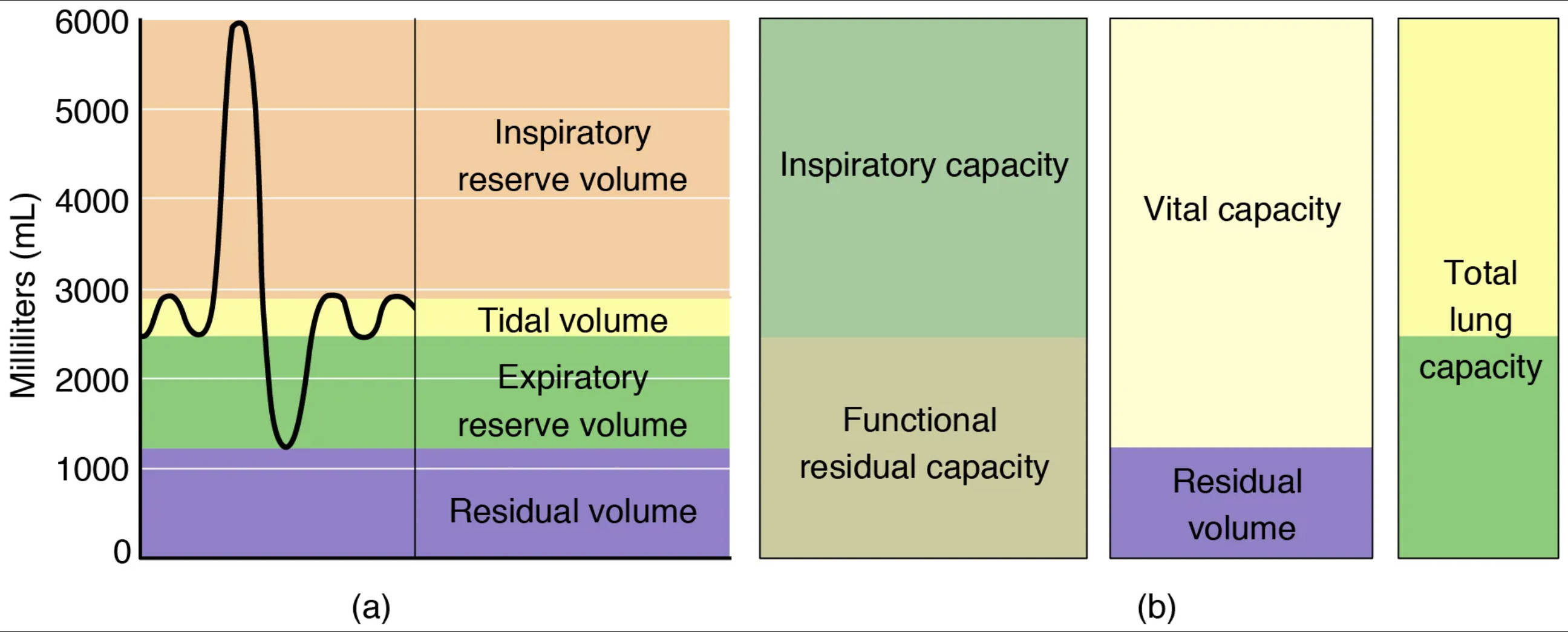
Inspiratory reserve volume: This represents the additional volume of air that can be forcibly inhaled after a normal inspiration. It is a measure of the lung’s ability to take in extra air beyond the usual tidal breath when needed, such as during exercise.
Tidal volume: This is the volume of air inhaled or exhaled during a single, normal, quiet breath. It reflects the amount of air exchanged during routine breathing and is a fundamental measure of respiratory function at rest.
Expiratory reserve volume: This refers to the additional volume of air that can be forcibly exhaled after a normal expiration. It represents the extra air that can be expelled from the lungs beyond the usual amount exhaled during quiet breathing.
Residual volume: This is the volume of air that remains in the lungs even after a maximal forceful expiration. This air can never be completely exhaled and is crucial for keeping the alveoli open and preventing lung collapse.
Inspiratory capacity: This is the maximum volume of air that can be inhaled after a normal expiration. It is calculated as the sum of the tidal volume and the inspiratory reserve volume, representing the total amount of air one can inspire from a resting expiratory level.
Functional residual capacity: This represents the volume of air remaining in the lungs after a normal, quiet expiration. It is the sum of the expiratory reserve volume and the residual volume, indicating the amount of air available for gas exchange between breaths.
Vital capacity: This is the maximum volume of air that can be exhaled after a maximal inspiration. It is a critical measure of overall lung function, encompassing the tidal volume, inspiratory reserve volume, and expiratory reserve volume.
Total lung capacity: This is the maximum volume of air that the lungs can hold after a maximal inspiration. It represents the sum of all four primary lung volumes (tidal, inspiratory reserve, expiratory reserve, and residual volumes), indicating the total storage capacity of the lungs.
The Foundation of Respiratory Assessment
Respiratory volumes and capacities are fundamental measurements used to assess lung function and diagnose various respiratory conditions. These metrics quantify the amount of air moved in and out of the lungs, as well as the air remaining within them, providing a comprehensive picture of an individual’s pulmonary health. By evaluating these parameters, healthcare professionals can gain crucial insights into the efficiency of gas exchange and the mechanical properties of the respiratory system.
These measurements are typically obtained through spirometry, a common non-invasive test. Spirometry plays a vital role in identifying restrictive or obstructive lung diseases, which can significantly impact an individual’s quality of life. Understanding the definitions and interrelationships of these volumes and capacities is essential for both medical practitioners and patients seeking to comprehend their respiratory health.
Key measurements include:
- Volumes: Tidal volume, inspiratory reserve volume, expiratory reserve volume, and residual volume.
- Capacities: Combinations of two or more volumes that provide broader insights into lung function.
These parameters allow for a detailed analysis of how well the lungs are performing their primary role of oxygen intake and carbon dioxide expulsion.
Diving Deeper into Lung Function Metrics
The efficiency of our respiratory system is often quantified by measuring the various volumes of air that our lungs can accommodate and exchange. These measurements are crucial for understanding the mechanics of breathing and detecting abnormalities that might indicate respiratory distress or disease. Each volume and capacity provides a distinct piece of the puzzle, contributing to a holistic understanding of pulmonary mechanics.
Primary Lung Volumes Explained
The four primary lung volumes are the building blocks for all respiratory capacities. The tidal volume (TV), typically around 500 mL in a healthy adult, represents the amount of air inhaled or exhaled during normal, quiet breathing. This volume reflects the baseline respiratory effort. The inspiratory reserve volume (IRV) is the additional air that can be forcibly inhaled after a normal tidal inspiration, often around 3000 mL. It signifies the potential for deeper breaths. Conversely, the expiratory reserve volume (ERV) is the extra air that can be forcibly exhaled after a normal tidal expiration, usually about 1100 mL. Finally, the residual volume (RV), approximately 1200 mL, is the air that always remains in the lungs, even after the most forceful exhalation. This residual air prevents the collapse of the alveoli and ensures continuous gas exchange between breaths.
Respiratory Capacities: Combinations for Comprehensive Assessment
Respiratory capacities are combinations of two or more lung volumes, offering a more comprehensive assessment of lung function. Inspiratory capacity (IC) is the maximum amount of air that can be inhaled after a normal expiration (TV + IRV), typically around 3500 mL. This indicates the total inspiratory ability from a resting expiratory level. Functional residual capacity (FRC) is the volume of air remaining in the lungs after a normal expiration (ERV + RV), usually about 2300 mL. FRC is particularly important as it represents the equilibrium point of the respiratory system, where the elastic recoil of the lungs balances the outward recoil of the chest wall. Vital capacity (VC) is the maximum amount of air that can be exhaled after a maximal inspiration (IRV + TV + ERV), often around 4600 mL. It is a strong indicator of the strength of respiratory muscles and the elasticity of the lungs. Lastly, total lung capacity (TLC) is the total amount of air the lungs can hold after a maximal inspiration (IRV + TV + ERV + RV), approximately 5800 mL. TLC represents the maximum possible volume of air in the lungs and is a comprehensive measure of lung size and distensibility. Abnormalities in these volumes and capacities can indicate conditions like asthma, COPD, or restrictive lung diseases, necessitating further diagnostic investigation.
Conclusion
The meticulous measurement and interpretation of respiratory volumes and capacities are indispensable tools in modern medicine for evaluating pulmonary function. These parameters not only provide a quantitative snapshot of lung mechanics but also serve as crucial indicators for diagnosing, monitoring, and managing a wide array of respiratory diseases. A thorough understanding of tidal volume, reserve volumes, residual volume, and their combinations into various capacities empowers healthcare professionals to make informed decisions, ultimately enhancing patient care and improving respiratory health outcomes.

