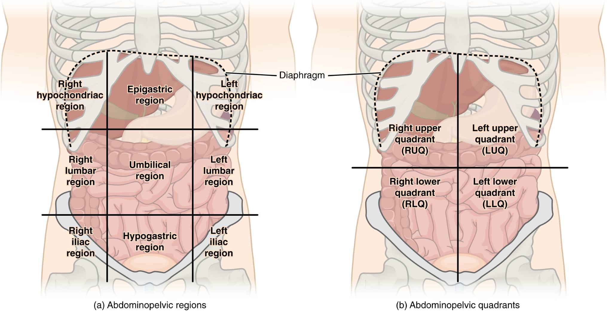
The peritoneal cavity is a vital space within the abdomen, housing numerous organs essential for digestion and metabolism. This image highlights the Right Hypochondriac Region, Epigastric Region, Left Hypochondriac Region, Right Lumbar Region, Umbilical Region, Left Lumbar Region, Right Iliac Region, Hypogastric Region, Left Iliac Region, Diaphragm, Right Upper Quadrant (RUQ), Left Upper Quadrant (LUQ), Right Lower Quadrant (RLQ), and Left Lower Quadrant (LLQ), offering a detailed view of both regional and quadrant divisions. Exploring these areas provides a clear framework for locating and studying the body’s internal structures with accuracy.
Label Introductions:
- Right Hypochondriac Region: The right hypochondriac region lies under the lower ribs on the right side, housing the liver and gallbladder. It is a key area for assessing upper abdominal pain or organ dysfunction.
- Epigastric Region: The epigastric region is centrally located above the stomach, containing the stomach and parts of the liver. It is often examined for issues like acid reflux or epigastric tenderness.
- Left Hypochondriac Region: The left hypochondriac region, under the left ribs, contains the spleen and part of the stomach. It is critical for evaluating spleen-related conditions or left-sided pain.
- Right Lumbar Region: The right lumbar region is situated in the middle right side, adjacent to the intestines. It is useful for diagnosing issues in the ascending colon or nearby structures.
- Umbilical Region: The umbilical region surrounds the navel, housing parts of the small intestine and transverse colon. It serves as a central point for assessing abdominal distension or pain.
- Left Lumbar Region: The left lumbar region, on the middle left side, includes the descending colon. It is important for identifying lower left abdominal discomfort or intestinal issues.
- Right Iliac Region: The right iliac region, in the lower right abdomen, contains the appendix. It is a focal point for diagnosing appendicitis or lower right pain.
- Hypogastric Region: The hypogastric region, or suprapubic area, lies below the navel and includes the bladder and reproductive organs. It is examined for urinary or pelvic conditions.
- Left Iliac Region: The left iliac region, in the lower left abdomen, houses parts of the sigmoid colon. It is relevant for assessing lower left quadrant pain or bowel irregularities.
- Diaphragm: The diaphragm separates the thoracic and abdominal cavities, aiding in breathing by contracting and relaxing. It plays a role in supporting abdominal organ positioning.
- Right Upper Quadrant (RUQ): The right upper quadrant includes the liver, gallbladder, and part of the stomach, located under the right rib cage. It is a common site for evaluating hepatobiliary disorders.
- Left Upper Quadrant (LUQ): The left upper quadrant contains the spleen, stomach, and pancreas, situated under the left rib cage. It is key for diagnosing splenic or gastric issues.
- Right Lower Quadrant (RLQ): The right lower quadrant houses the appendix and parts of the intestines, located in the lower right abdomen. It is critical for identifying appendicitis or bowel obstruction.
- Left Lower Quadrant (LLQ): The left lower quadrant includes the sigmoid colon and left ovary, positioned in the lower left abdomen. It is examined for conditions like diverticulitis or ovarian cysts.
Overview of the Peritoneal Cavity
The peritoneal cavity is a lined space within the abdomen, encompassing organs like the stomach, liver, and intestines. Divided into nine abdominopelvic regions and four quadrants, it provides a structured approach to anatomical study and clinical assessment. These divisions help in pinpointing the location of organs and diagnosing related conditions with precision.
- Protects and supports abdominal organs with a serous membrane.
- Facilitates movement of organs during digestion and respiration.
- Allows for targeted medical imaging and surgical planning.
- Serves as a reference for documenting abdominal pain or abnormalities.
Abdominopelvic Regions: Detailed Segmentation
The nine abdominopelvic regions offer a fine-grained division of the abdominal area. The right hypochondriac region and left hypochondriac region are upper zones near the ribs, while the umbilical region marks the central area. This detailed mapping aids in localizing specific organ-related symptoms.
- The epigastric region is above the stomach, often linked to ulcer pain.
- The right and left lumbar regions flank the umbilical area, housing intestinal loops.
- The hypogastric region supports the bladder, influencing urinary health.
- The right and left iliac regions are key for appendiceal and colonic assessments.
Abdominopelvic Quadrants: Broad Divisions
The four abdominopelvic quadrants simplify the abdominal layout into larger sections. The right upper quadrant (RUQ) includes the liver, while the left upper quadrant (LUQ) contains the spleen. These quadrants are widely used in emergency medicine for quick assessments.
- The right lower quadrant (RLQ) is a primary site for appendicitis evaluation.
- The left lower quadrant (LLQ) is checked for diverticular disease.
- The diaphragm separates the upper quadrants from the thoracic cavity.
- Quadrants guide initial physical exams and diagnostic imaging.
Role of the Diaphragm
The diaphragm is a muscular structure that divides the thoracic and abdominal cavities. It plays a crucial role in respiration by moving downward to allow lung expansion. Its position also influences the placement of upper abdominal organs.
- Contracts during inhalation to increase thoracic volume.
- Relaxes during exhalation to push air out.
- Supports the liver and stomach in the upper quadrants.
- Can be affected by conditions like hiatal hernia.
Clinical Relevance of Regions and Quadrants
Understanding the abdominopelvic regions and quadrants is essential for clinical practice. The right iliac region is a common site for appendicitis, while the epigastric region may indicate gastric ulcers. These divisions enhance the accuracy of physical exams and imaging interpretations.
- The RUQ is assessed for gallbladder inflammation or gallstones.
- The LUQ helps diagnose splenomegaly or pancreatic issues.
- The RLQ is critical for detecting ovarian torsion in females.
- The LLQ aids in identifying sigmoid colon disorders.
Conclusion
The exploration of the abdominopelvic regions and quadrants within the peritoneal cavity reveals the intricate organization of abdominal anatomy. From the right hypochondriac region to the left lower quadrant (LLQ), each area plays a unique role in housing and protecting vital organs. This knowledge equips professionals to navigate the abdomen effectively, improving diagnostic and therapeutic outcomes in medical care.

