The human body’s ability to perceive and respond to the environment hinges on the diverse structures of sensory receptors, which are classified based on their cellular composition. These receptors, depicted in this image, include neurons with free nerve endings, encapsulated nerve endings, and specialized cells like photoreceptors, each playing a unique role in sensory processing. This article provides an in-depth exploration of these receptor types, their anatomical features, and their critical functions in translating external stimuli into neural signals.
Labeled Parts of Receptor Classification
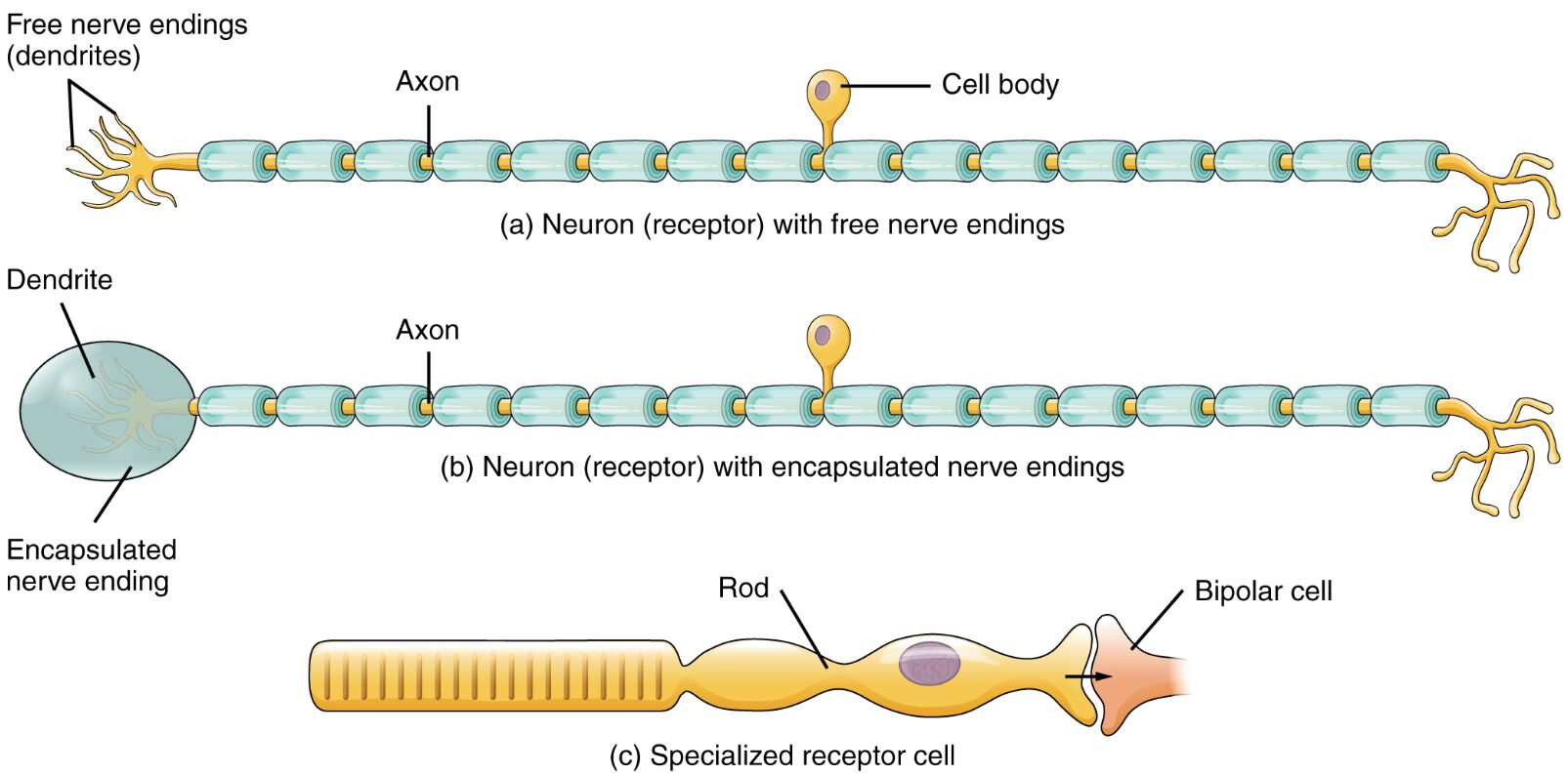
Free nerve endings (dendrites) Free nerve endings are unmyelinated or lightly myelinated nerve terminals that serve as receptors for pain, temperature, and crude touch. They extend from the dendrites of sensory neurons and are widely distributed in the skin, mucous membranes, and other tissues, making them essential for detecting potentially harmful stimuli.
Axon The axon is the long, slender projection of a neuron that conducts electrical impulses away from the cell body toward other neurons or effector cells. In sensory neurons, it transmits sensory information from the receptor site to the central nervous system, playing a key role in signal propagation.
Cell body The cell body, or soma, contains the nucleus and maintains the neuron’s metabolic functions, supporting the synthesis of proteins and neurotransmitters. It serves as the control center for the neuron, integrating incoming signals before they are relayed along the axon.
Dendrite Dendrites are branched extensions of the neuron that receive incoming signals from other neurons or sensory stimuli, funneling them toward the cell body. In receptors with encapsulated endings, dendrites are enclosed within specialized structures, enhancing their sensitivity to specific stimuli.
Encapsulated nerve ending Encapsulated nerve endings are sensory receptors surrounded by connective tissue or other cellular structures, designed to detect pressure, vibration, or stretch. Examples include Meissner’s corpuscles and Pacinian corpuscles, which provide detailed tactile and proprioceptive information.
Rod The rod is a specialized photoreceptor cell in the retina of the eye, highly sensitive to low light and responsible for night vision and peripheral vision. It contains rhodopsin, a pigment that undergoes photochemical changes to initiate visual signal transduction.
Bipolar cell The bipolar cell is a type of neuron in the retina that connects photoreceptors like rods to ganglion cells, facilitating the transmission of visual information. It integrates signals from multiple photoreceptors and synapses with the optic nerve, playing a critical role in visual processing.
Anatomical Structure of Sensory Receptors
Sensory receptors are specialized structures that convert external stimuli into electrical signals, initiating the sensory process. Their classification into different cell types reflects their adaptation to specific sensory modalities.
- Free nerve endings: These receptors lack a protective capsule, allowing them to respond to a broad range of stimuli, including pain and temperature changes, across various tissues.
- Encapsulated endings: The surrounding capsule enhances sensitivity to mechanical stimuli, with structures like Pacinian corpuscles detecting deep pressure and vibration.
- Specialized cells: Photoreceptors like rods are highly differentiated, located in the retina, and optimized for low-light conditions, contributing to the eye’s adaptability.
- Neuronal components: The axon and dendrites work in tandem with the cell body, ensuring efficient signal transmission from peripheral receptors to the central nervous system.
- Retinal integration: In the visual system, rods synapse with bipolar cells, forming a complex network that processes light into actionable neural signals.
Physiological Functions of Receptor Types
Each receptor type is tailored to detect specific stimuli, contributing to the body’s sensory landscape. Their physiological roles are critical for survival and interaction with the environment.
- Pain and temperature: Free nerve endings activate nociceptors and thermoreceptors, alerting the body to potential injury or thermal extremes.
- Tactile sensation: Encapsulated endings, such as Meissner’s corpuscles, provide fine touch and vibration detection, essential for manipulating objects.
- Vision: Rods enable low-light vision by converting photons into electrical signals, while bipolar cells relay this information to the optic nerve for further processing.
- Signal transduction: The axon conducts action potentials, while dendrites receive initial stimuli, ensuring a seamless flow of sensory data.
- Adaptation mechanisms: Receptors adapt to sustained stimuli, with encapsulated endings adjusting sensitivity to maintain relevant sensory input.
Developmental and Cellular Organization
The development of sensory receptors involves intricate cellular differentiation, shaping their structure and function during embryogenesis. This process ensures that each receptor type aligns with its sensory role.
- Neural crest origins: Free nerve endings and encapsulated endings derive from neural crest cells, which migrate to form peripheral sensory structures.
- Retinal development: Rods and bipolar cells develop from the optic vesicle, with rods specializing for scotopic vision and bipolar cells forming synaptic connections.
- Myelination: Axons may be myelinated to enhance signal speed, with unmyelinated free nerve endings relying on rapid depolarization for response.
- Cellular synergy: The cell body supports dendritic growth and axonal elongation, coordinating the neuron’s sensory and conductive roles.
- Evolutionary adaptations: Specialized receptors like rods reflect evolutionary pressures for enhanced night vision in early vertebrates.
Clinical Relevance and Sensory Disorders
Understanding receptor classification aids in diagnosing and managing sensory-related conditions. Clinical assessments often focus on the integrity of these structures to pinpoint dysfunction.
- Neuropathy: Damage to free nerve endings can lead to loss of pain sensation, increasing injury risk in conditions like diabetes.
- Vision impairment: Rod dysfunction, as in retinitis pigmentosa, causes night blindness due to impaired photoreceptor function.
- Tactile deficits: Encapsulated ending disorders may result in reduced vibration sense, affecting coordination and balance.
- Diagnostic tools: Electroretinography assesses rod and bipolar cell activity, while nerve conduction studies evaluate axonal function.
- Therapeutic strategies: Treatments range from vitamin A supplementation for rod health to nerve repair for axonal damage.
In conclusion, the classification of sensory receptors by cell type highlights the remarkable diversity and specialization within the nervous system. From free nerve endings detecting pain to rods enabling night vision, these structures form the foundation of sensory perception, offering valuable insights into both normal physiology and potential pathologies.

