Photoreceptors are the specialized cells within the retina that capture light and initiate the process of vision, making them essential to our ability to see. This image provides a detailed look at the anatomical structure of these cells, highlighting the differences between rods and cones and their critical components.
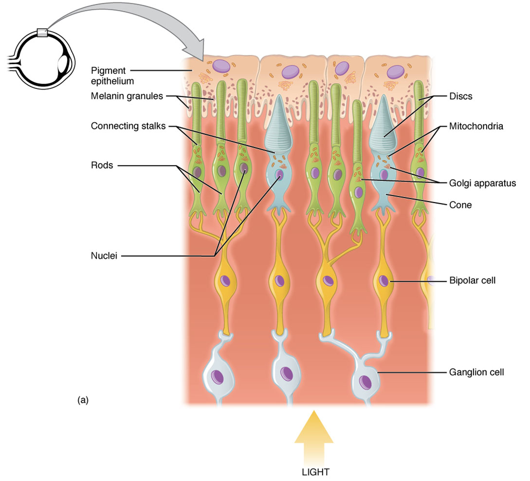
Inner segment The inner segment contains the nucleus and vital organelles, serving as the metabolic hub of photoreceptors. It connects to the outer segment, supplying the energy needed for light detection.
Outer segment The outer segment is packed with membrane arrays containing photosensitive opsin molecules, where light absorption occurs. In rods, it forms long columnar shapes with stacked discs holding rhodopsin, while in cones, it features short, tapered shapes with membrane folds.
Rod The rod is a photoreceptor designed for low-light vision, characterized by its long outer segment filled with rhodopsin pigment. Its high sensitivity allows it to function effectively in dim conditions but offers limited color perception.
Cone The cone is a photoreceptor optimized for color and high-acuity vision, with a short, tapered outer segment. It contains opsins sensitive to red, green, or blue light, performing best in bright environments.
Anatomy of Photoreceptors
Photoreceptors are the retina’s light-sensitive cells, divided into distinct segments that support their function. This image showcases the structural differences that enable rods and cones to handle diverse lighting conditions.
- The inner segment houses mitochondria, providing ATP for photoreceptor activity.
- The outer segment contains the photopigments critical for converting light into electrical signals.
- Rods have a cylindrical shape, with thousands of membrane discs increasing rhodopsin exposure.
- Cones are shorter and tapered, with folded membranes that allow quicker photopigment renewal.
- The connection between inner and outer segments is maintained by a cilium, ensuring structural integrity.
- Supporting cells, such as Müller cells, provide nutrients and structural support to these photoreceptors.
- The retina’s layered arrangement positions these cells optimally for light capture.
Physiology of Photoreceptor Function
Photoreceptors transform light into neural signals through a sophisticated process tailored to their structure. The distinct designs of rods and cones underpin their specialized visual roles.
- The outer segment of rods maximizes rhodopsin concentration, enhancing sensitivity in low light.
- Cones have a more compact outer segment, supporting faster response times for color detection.
- Light absorption by opsin in the outer segment triggers a biochemical cascade, hyperpolarizing the cell.
- Rods detect single photons, making them ideal for night vision, but they saturate in bright light.
- Cones require higher light levels, enabling sharp vision and color differentiation.
- The inner segment ensures continuous energy supply, sustaining this phototransduction process.
- Signals are relayed to bipolar cells, initiating the visual pathway to the brain.
Role of Rods in Low-Light Vision
Rods are engineered for exceptional sensitivity, thriving in dim environments. Their elongated structure enhances their ability to capture light efficiently.
- The long outer segment of rods contains stacked discs, amplifying rhodopsin’s light-absorbing capacity.
- Rhodopsin, a light-sensitive pigment, breaks down into retinal and opsin upon photon absorption.
- This breakdown initiates a signal amplification process, detectable even in near darkness.
- Rods lack color sensitivity, relying on a single photopigment type for monochromatic vision.
- Their prevalence in the retina’s periphery supports wide-field, low-acuity vision.
- Dark adaptation involves rhodopsin regeneration, a process that can take up to 30 minutes.
Role of Cones in Color and Detail Vision
Cones are tailored for bright light, with a structure that supports color perception and high resolution. Their design makes them essential for daytime visual tasks.
- The tapered outer segment of cones features folded membranes, allowing rapid opsin renewal.
- Three cone types—L, M, and S—contain opsins for red, green, and blue light detection.
- This trichromatic system enables color vision through neural comparison in the brain.
- Cones are densely packed in the fovea, providing the retina’s highest visual acuity.
- Their lower sensitivity requires ample light, complementing rods in bright conditions.
- Cone dysfunction can lead to color blindness, though this image depicts normal anatomy.
Supporting Structures and Cellular Integration
The inner segment and outer segment work together, supported by surrounding retinal cells. This integration ensures efficient photoreceptor performance.
- The inner segment contains the endoplasmic reticulum, synthesizing photopigments.
- The cilium linking segments maintains structural continuity and signal transmission.
- Müller cells provide structural support and remove metabolic waste from photoreceptors.
- Horizontal and amacrine cells modulate signals from rods and cones in the outer plexiform layer.
- The retinal pigment epithelium nourishes the outer segment, recycling photopigments.
- This cellular network enhances the retina’s ability to adapt to varying light levels.
Clinical Relevance of Photoreceptors
Knowledge of photoreceptor anatomy assists in identifying and managing vision impairments. This image serves as a reference for understanding normal structure and function.
- Retinitis pigmentosa targets rods initially, leading to night blindness and progressive vision loss.
- Age-related macular degeneration affects cones in the fovea, impairing central vision.
- Damage to the outer segment can result from oxidative stress, a factor in retinal diseases.
- Cone deficiencies may cause color vision disorders, such as deuteranopia.
- The inner segment’s metabolic health is crucial, as mitochondrial dysfunction can harm photoreceptors.
- Treatments like vitamin A therapy support rod function in early disease stages.
- Advanced imaging techniques monitor photoreceptor integrity for timely interventions.
In conclusion, the photoreceptors of the eye, as illustrated in this anatomical view, demonstrate a remarkable adaptation for vision across different light conditions. Their intricate structure and specialized functions highlight the eye’s ability to process a wide range of visual information, making this a compelling area for exploring ocular health.

