The bacterial cell wall is a complex and essential structure that provides physical protection and maintains cellular shape. Peptidoglycan, a polymer of sugars and amino acids, forms a mesh-like layer that varies significantly between Gram-positive and Gram-negative bacteria. Understanding the molecular arrangement of these components is vital for medical professionals in the diagnosis and treatment of bacterial infections.
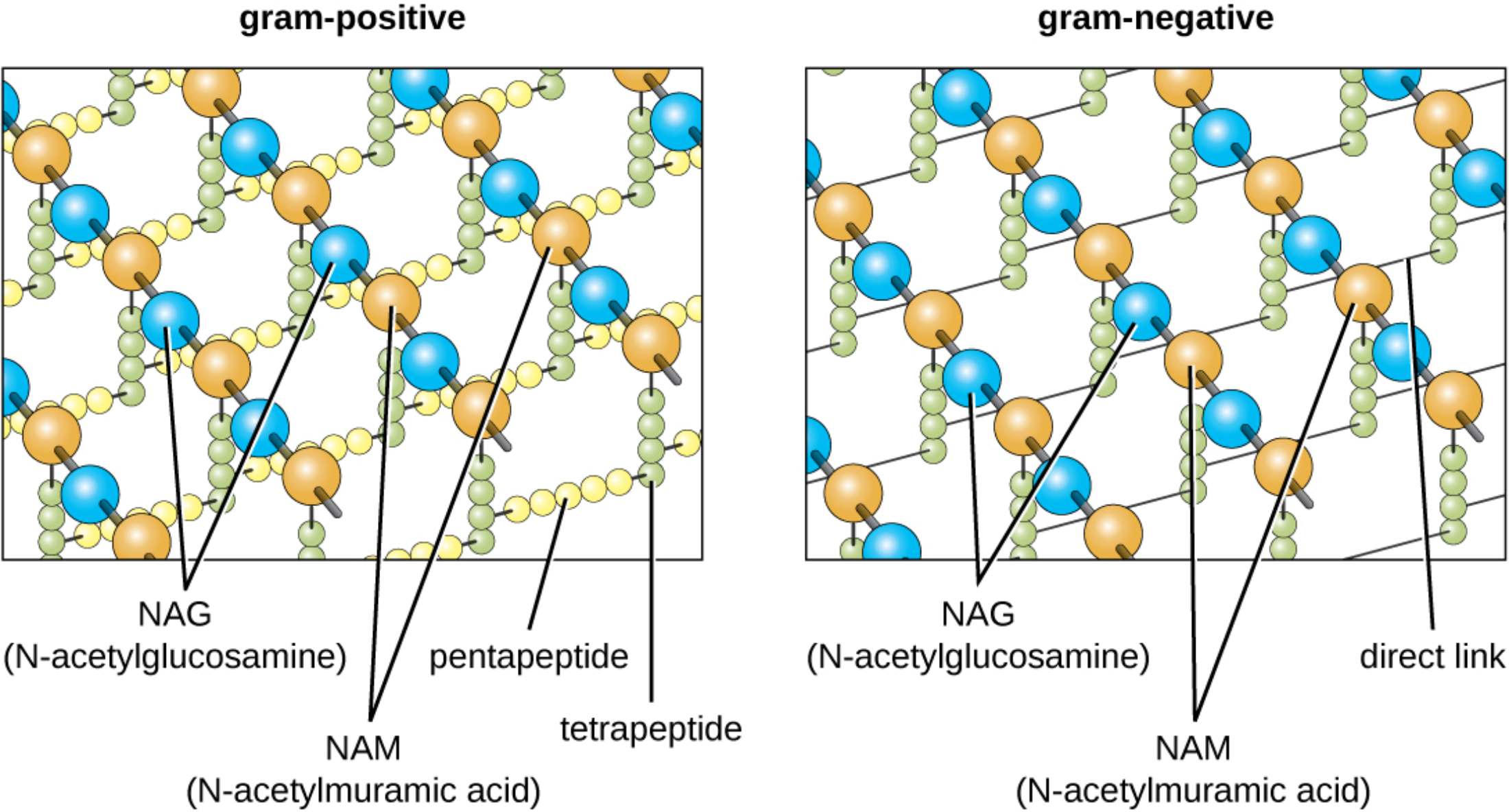
gram-positive: This term refers to bacteria that possess a thick layer of peptidoglycan in their cell wall, which retains the crystal violet stain used in laboratory testing. This structural density provides significant protection against environmental stressors but leaves the bacteria vulnerable to certain classes of antibiotics.
gram-negative: These organisms are characterized by a thinner peptidoglycan layer and an additional outer membrane containing lipopolysaccharides. This unique architecture acts as a barrier to many common antibiotics and prevents the retention of primary stains during Gram staining procedures.
NAG (N-acetylglucosamine): This is one of the two alternating amino sugars that form the glycan strands of the bacterial cell wall. It provides a structural foundation that, when linked with NAM, creates the rigid backbone necessary for maintaining cellular integrity.
NAM (N-acetylmuramic acid): This sugar molecule alternates with NAG to build the carbohydrate chain and serves as the attachment site for peptide side chains. The specific chemical bonds formed at this subunit are critical for the cross-linking that gives the cell wall its tensile strength.
pentapeptide: Found commonly in Gram-positive bacteria, this chain of five amino acids forms a bridge that connects parallel glycan strands. This bridge adds a layer of flexibility and thickness to the cell wall, contributing to its overall resilience.
tetrapeptide: This is a chain of four amino acids that extends from the NAM subunits in the peptidoglycan structure. It plays a fundamental role in the cross-linking process, allowing various glycan chains to bind together into a cohesive network.
direct link: In many Gram-negative bacteria, a direct covalent bond forms between the peptide side chains of adjacent glycan strands. This configuration results in a more compact and rigid lattice structure compared to the interbridge found in other bacterial types.
The Architecture of Microbial Survival
The architecture of the bacterial cell wall is one of the most studied topics in microbiology due to its fundamental role in survival. Peptidoglycan, a complex macromolecule unique to bacteria, functions as a protective “cage” that prevents the cell from bursting under high internal osmotic pressure. This structure is so vital that interfering with its synthesis is one of the most effective strategies for modern medical therapy.
While the basic building blocks remain consistent across many species, the spatial arrangement of these molecules creates the defining characteristics used to classify bacteria. The distinction between Gram-positive and Gram-negative organisms is based primarily on the thickness and connectivity of the peptidoglycan layer. This differentiation influences not only how bacteria look under a microscope but also how they respond to the human immune system and pharmaceutical interventions.
Important features of the peptidoglycan structure include:
- Alternating NAG and NAM units forming a linear glycan backbone.
- Horizontal stability provided by beta-1,4-glycosidic bonds.
- Vertical and lateral stability established through peptide side chains.
- Specialized amino acids, such as D-alanine, that are resistant to most proteases.
By analyzing these microscopic details, clinicians can better understand the mechanism of action for various treatments. The presence of specialized bridges or direct links impacts the porosity of the cell wall, affecting the diffusion of nutrients and the entry of antimicrobial agents.
The Glycan Backbone and Molecular Integrity
The linear chains of N-acetylglucosamine (NAG) and N-acetylmuramic acid (NAM) provide the longitudinal strength of the cell wall. These sugars are joined by specialized bonds that are remarkably stable, yet dynamic enough to allow for cell growth and division. In the medical field, the integrity of this backbone is a primary target; for example, human lysozyme functions by cleaving these sugar bonds, effectively dissolving the bacterial shield in fluids like tears and saliva.
Peptide Connectivity and Antibiotic Action
The lateral stability of the cell wall is maintained through cross-linking between peptide chains. In Gram-positive bacteria, a pentapeptide bridge often provides the necessary spacing between strands, whereas Gram-negative bacteria utilize a direct link for a more streamlined profile. This difference in connectivity is a critical factor in how various medications exert their effects on different pathogens.
For instance, beta-lactam antibiotics, such as penicillin and cephalosporins, work by inhibiting the transpeptidase enzymes responsible for forming these links. When these enzymes are blocked, the cell wall becomes mechanically unstable, leading to bacterial death via lysis. Understanding these molecular nuances helps healthcare providers select the most appropriate narrow-spectrum or broad-spectrum treatments based on the suspected bacterial structure.
The intricate design of peptidoglycan illustrates the complexity of microbial life. From the alternating sugar subunits to the specialized peptide bridges, every element is optimized for survival in diverse environments. By targeting these unique structures, modern medicine continues to refine therapies that protect human health while respecting the biological differences between host cells and invading pathogens.

