Explore the remarkable journey of blood as it circulates through the human heart, a process fundamental to life, as clearly illustrated in this diagram. This explanation will detail the precise pathway of both deoxygenated and oxygenated blood, highlighting how the heart efficiently pumps nutrients and oxygen throughout the body. A comprehensive understanding of normal blood flow is essential for recognizing deviations and potential cardiovascular issues.
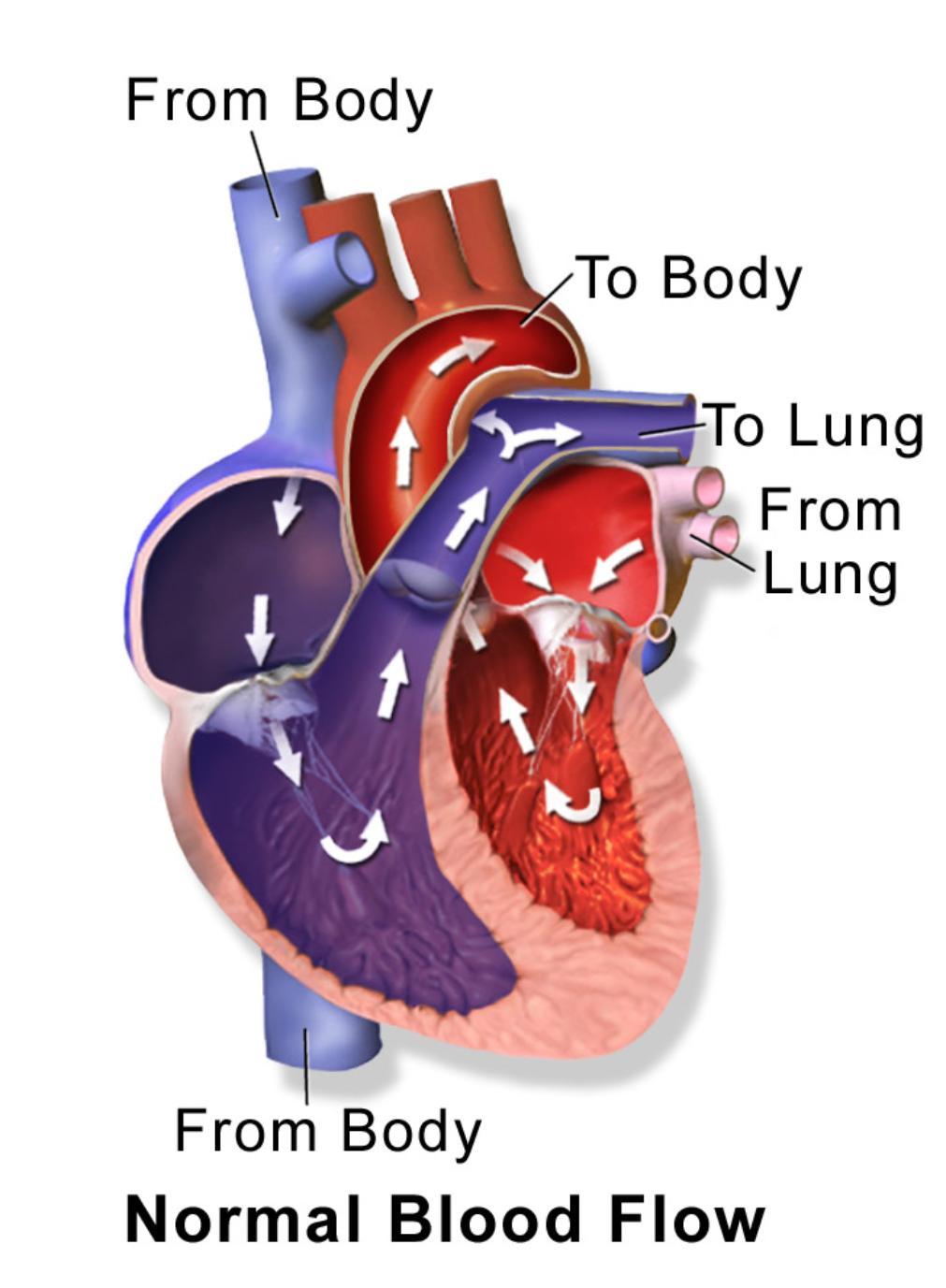
From Body: This label indicates the entry point of deoxygenated blood returning from the systemic circulation, specifically through the superior and inferior vena cava. This blood is rich in carbon dioxide and metabolic waste products, requiring re-oxygenation.
To Body: This label signifies the exit point for oxygenated blood leaving the heart to be distributed to the rest of the body via the aorta. This blood is rich in oxygen and nutrients, essential for cellular function.
To Lung: This label points to the pathway of deoxygenated blood being pumped from the heart to the lungs through the pulmonary artery. In the lungs, carbon dioxide is released, and oxygen is picked up.
From Lung: This label denotes the entry point of oxygenated blood returning from the lungs to the heart via the pulmonary veins. This newly oxygenated blood is now ready to be pumped to the rest of the body.
The human heart is a complex, four-chambered organ that acts as a tireless pump, driving blood through two distinct circulatory pathways: the pulmonary circulation and the systemic circulation. This diagram vividly illustrates the seamless, unidirectional flow of blood, a process vital for delivering oxygen and nutrients to every cell while simultaneously removing waste products. Understanding this normal blood flow pattern is the cornerstone of comprehending cardiovascular physiology and is crucial for identifying when and where abnormalities might occur.
The journey begins with deoxygenated blood (depicted in blue/purple) returning From Body tissues, entering the heart’s right atrium. From there, it passes into the right ventricle, which then pumps it To Lung through the pulmonary artery. In the lungs, a vital exchange occurs: carbon dioxide is released, and oxygen is absorbed. The newly oxygenated blood (depicted in red) then travels From Lung back to the heart, entering the left atrium.
From the left atrium, the oxygenated blood flows into the powerful left ventricle, which then forcefully pumps it To Body through the aorta, distributing it to all tissues and organs. This continuous cycle ensures that every part of the body receives the necessary resources to function. The heart’s valves play a critical role in this process, acting as one-way gates to prevent the backflow of blood, thus maintaining the efficiency of this life-sustaining circulation. Any disruption to this normal flow, such as a leaky valve or a narrowed vessel, can significantly impact overall health.
- The heart beats approximately 100,000 times a day, circulating about 5 liters of blood per minute.
- The pulmonary circulation is a low-pressure system, while the systemic circulation is a high-pressure system.
- The right side of the heart handles deoxygenated blood, and the left side handles oxygenated blood.
- The entire blood volume circulates through the body in about one minute.
Understanding the normal blood flow through the heart is fundamental not only for medical professionals but also for individuals seeking to grasp the basics of their own health. Conditions like congenital heart defects, which can involve abnormal openings between chambers or narrowed vessels, directly interfere with this flow, potentially leading to oxygen deprivation in tissues or excessive workload on the heart. Knowledge of this pathway empowers us to appreciate the heart’s incredible work and to recognize the importance of maintaining cardiovascular health.
This diagram beautifully simplifies the complex yet essential process of blood flow through the heart, making it accessible and easy to understand. The continuous, coordinated movement of blood, driven by the heart’s pumping action and guided by its internal structures, is a fundamental aspect of human physiology. Grasping this normal pathway is key to appreciating the heart’s vital role in sustaining life and to identifying deviations that may indicate cardiovascular health issues.

