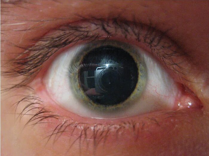
The human eye offers a fascinating window into the autonomic nervous system’s influence, with pupil dilation, or mydriasis, serving as a key indicator of physiological responses. This image captures a striking example of mydriasis, where the pupil appears significantly enlarged due to the activation of sympathetic pathways or the application of specific medications like phenylephrine. Delving into this visual provides valuable insights into the eye’s anatomy and the mechanisms behind its involuntary adjustments, making it an essential topic for anyone interested in ocular health.
Iris The iris is the colored part of the eye surrounding the pupil, controlling its size through smooth muscle contractions. It contains radial and circular muscles that respond to autonomic signals, enabling the pupil to adjust light entry and focus.
Pupil The pupil is the central opening in the iris that regulates light entering the eye, dilating or constricting based on environmental conditions or nervous system activity. In this image, its enlarged state reflects mydriasis, likely induced by sympathetic stimulation or pharmacological agents.
Eyelashes Eyelashes are protective hairs along the eyelid’s edge, shielding the eye from debris and reducing moisture loss. They frame the eye in this image, highlighting the dilated pupil’s prominence against the surrounding structures.
Sclera The sclera is the white, tough outer layer of the eyeball, providing structural support and protecting the inner eye components. It appears prominently in the image, contrasting with the dilated pupil and colored iris.
Anatomy of the Eye and the Role of the Iris
The eye’s intricate design allows for dynamic responses to light and internal signals. The iris plays a central role in this process, adapting the pupil’s size accordingly.
- The iris contains two muscle groups: the radial dilator muscles and the circular sphincter muscles.
- Radial muscles, innervated by the sympathetic nervous system, contract to cause pupil dilation during low-light conditions or stress.
- The circular muscles, controlled by the parasympathetic system via the oculomotor nerve, constrict the pupil in bright light.
- This dual control ensures optimal vision, with the pupil adjusting from a tiny pinhole to a wide opening as seen in mydriasis.
- Blood supply to the iris, via the anterior ciliary arteries, supports these rapid muscular responses.
Mechanisms Behind Pupil Dilation (Mydriasis)
Mydriasis occurs when the pupil enlarges, often due to sympathetic activation or medical intervention. This image showcases the effect, offering a clear view of the process.
- The sympathetic nervous system releases norepinephrine, binding to alpha-1 adrenergic receptors on the iris’s radial fibers.
- This binding triggers muscle contraction, pulling the iris outward to widen the pupil, as seen in the photograph.
- Phenylephrine, a synthetic agonist, mimics this action by activating the same receptors when applied as eye drops.
- Such dilation is useful in ophthalmology for examining the retina or managing certain eye conditions.
- Prolonged dilation without medical supervision can lead to light sensitivity or discomfort due to uncontrolled light exposure.
Physiological Triggers of Sympathetic Activation
The sympathetic system’s role extends beyond the eye, influencing various bodily functions during stress or excitement. Pupil dilation is a visible sign of this response.
- Activation begins in the hypothalamus, sending signals through the spinal cord to the superior cervical ganglion.
- Postganglionic fibers then innervate the iris, releasing norepinephrine to induce mydriasis.
- This response prepares the body for action, enhancing visual acuity in dim environments.
- Hormones like adrenaline, released by the adrenal medulla, amplify this effect, supporting fight-or-flight readiness.
- Thyroid hormones such as T3 and T4 can increase metabolic demand, indirectly affecting pupil responsiveness.
Clinical Applications of Phenylephrine-Induced Mydriasis
Phenylephrine is a common tool in medical settings to achieve controlled pupil dilation. This image reflects its effective use in practice.
- Applied as eye drops, phenylephrine selectively stimulates alpha-1 receptors, bypassing natural triggers.
- This allows ophthalmologists to perform detailed retinal exams by widening the pupil for better visualization.
- The drug’s rapid onset and short duration make it ideal for diagnostic procedures lasting 30-60 minutes.
- Care must be taken to monitor intraocular pressure, as excessive dilation can strain the eye in glaucoma patients.
- Patients may experience temporary blurred vision, necessitating guidance post-examination.
Potential Risks and Considerations
While mydriasis can be beneficial, it carries risks if not managed properly. This image prompts a closer look at these concerns.
- Unintentional dilation from systemic sympathomimetic drugs can lead to photophobia or eye strain.
- In rare cases, prolonged mydriasis may signal underlying neurological issues, such as third nerve palsy.
- Individuals with narrow-angle glaucoma face heightened risk of acute angle-closure, requiring immediate attention.
- Overuse of dilating agents like phenylephrine can desensitize receptors, reducing future effectiveness.
- Regular eye check-ups help identify when dilation is a symptom rather than a treatment effect.
Normal vs. Pathological Mydriasis
Distinguishing between normal and abnormal pupil dilation is crucial for health assessment. This image provides a baseline for comparison.
- Normal mydriasis occurs in dim light or during emotional arousal, regulated by the autonomic nervous system.
- Pathological mydriasis may result from trauma, drug overdose, or brain injury, often unilateral and unresponsive to light.
- Anisocoria, unequal pupil sizes, can indicate nerve damage or inflammation affecting the iris.
- Monitoring pupil response with a penlight helps differentiate these states in clinical settings.
- Persistent dilation without cause warrants neuroimaging to rule out intracranial pathology.
Maintaining Eye Health Amid Autonomic Changes
Protecting eye health involves understanding how autonomic changes affect the iris and pupil. This image underscores the need for awareness.
- Wearing sunglasses can mitigate discomfort from dilated pupils in bright conditions.
- Regular hydration supports tear production, easing strain on the sclera and surrounding tissues.
- Avoiding stimulants like caffeine may reduce unnecessary sympathetic activation.
- Annual eye exams ensure the iris and pupil respond appropriately to light and medication.
- Balanced nutrition, rich in vitamin A and omega-3s, supports overall ocular function.
In conclusion, this image of mydriasis offers a compelling glimpse into the eye’s autonomic regulation, highlighting the interplay between the sympathetic nervous system and pharmacological interventions like phenylephrine. By exploring the anatomy, mechanisms, and clinical implications, we gain a deeper appreciation for the eye’s adaptability and the importance of professional oversight in managing pupil dilation. Whether for diagnostic purposes or understanding physiological responses, this visual serves as a valuable educational tool, encouraging proactive care to maintain optimal vision and eye health.

