Feline Aortic Thromboembolism (FATE), commonly known as a saddle thrombus, is a devastating condition where a blood clot obstructs the distal aorta in cats. This article provides a detailed anatomical and pathological insight into this critical veterinary emergency, explaining the location and impact of the thrombus on blood flow to the hind limbs and other vital organs. Discover the crucial vascular structures involved and the severe consequences of this life-threatening condition.
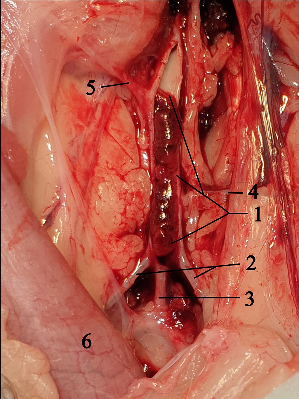
1 Opened Aorta with thrombus: This label points to the main artery, the aorta, which has been surgically opened to reveal a large blood clot, or thrombus, obstructing its lumen. This obstruction severely impedes blood flow to the posterior regions of the body, including the hind limbs.
2 A. iliaca externa (External Iliac Artery): This artery branches off the aorta and is responsible for supplying blood to the hind limbs. Its partial or complete blockage due to the saddle thrombus leads to the characteristic clinical signs observed in FATE, such as paralysis and pain in the rear limbs.
3 Common trunk for both Aa. iliacae internae (Internal Iliac Arteries): This refers to the shared origin for the internal iliac arteries, which branch to supply blood to the pelvic organs, gluteal muscles, and perineum. While often less severely affected than the external iliac arteries, their compromised blood supply contributes to the overall systemic impact of a saddle thrombus.
4 A. circumflexa ilium profunda (Deep Circumflex Iliac Artery): This artery typically originates near the iliac arteries and supplies blood to the muscles and skin of the flank region. Its involvement in the thrombotic event further illustrates the extensive vascular compromise in cases of saddle thrombus.
5 A. mesenterica caudalis (Caudal Mesenteric Artery): This artery branches off the aorta to supply blood to the descending colon and rectum. While not directly occluded by the saddle thrombus in its primary location, reduced aortic flow can still indirectly affect its perfusion and the viability of the caudal intestines.
6 Colon descendens (Descending Colon): This is the final segment of the large intestine, responsible for forming and storing feces before elimination. Its proximity to the affected aortic region highlights the potential for broader abdominal vascular compromise in severe cases of FATE.
Feline Aortic Thromboembolism (FATE), often referred to as a “saddle thrombus” due to its typical location at the aortic trifurcation (where the aorta divides into the iliac arteries), is an acute and severe condition in cats. It occurs when a blood clot, typically originating from the heart, dislodges and travels downstream until it becomes lodged in a narrower vessel, most commonly at the point where the abdominal aorta splits to supply the hind limbs. This sudden and critical obstruction of blood flow leads to severe pain, paralysis, and coldness in the affected hind limbs, constituting a medical emergency.
The image provided offers a stark visual representation of a saddle thrombus in the feline aorta, revealing the macroscopic pathology that underlies this devastating condition. By dissecting and opening the aorta, the large blood clot is clearly visible, dramatically illustrating how completely it can occlude the vessel lumen. This severe blockage immediately cuts off the oxygen and nutrient supply to the tissues distal to the thrombus, particularly the powerful muscles of the hind limbs, leading to rapid tissue damage and dysfunction.
Understanding the precise anatomical location of the thrombus and the arteries it affects is crucial for comprehending the clinical signs and guiding emergency treatment for FATE. The image highlights the critical branches of the aorta, such as the external iliac arteries, which are directly responsible for supplying the hind limbs. Compromise to these vessels results in the characteristic clinical presentation of acute hind limb paresis or paralysis, intense pain, and cold extremities, demanding immediate veterinary intervention to attempt to restore perfusion and alleviate suffering.
The majority of feline aortic thromboembolism cases are a sequela to underlying heart disease, particularly hypertrophic cardiomyopathy. This makes FATE not just a vascular emergency, but also a strong indicator of significant underlying cardiac pathology.
- Primary cause: Often linked to underlying heart disease, such as hypertrophic cardiomyopathy.
- Location: Typically at the aortic trifurcation, blocking hind limb blood supply.
- Clinical signs: Acute hind limb pain, paralysis, and coldness.
- Prognosis: Often guarded, necessitating rapid diagnosis and intervention.
This detailed anatomical view serves as a powerful educational tool for veterinary professionals and provides critical context for pet owners facing this challenging diagnosis.
Pathogenesis and Underlying Causes of FATE
The pathogenesis of Feline Aortic Thromboembolism (FATE) is primarily rooted in underlying cardiac disease, with hypertrophic cardiomyopathy (HCM) being the most common culprit. In HCM, the heart muscle thickens, particularly the walls of the left ventricle, which can lead to inefficient pumping and turbulent blood flow within the heart chambers. This abnormal blood flow, coupled with the enlargement of the left atrium and potential endothelial damage within the heart, creates an environment conducive to thrombus (blood clot) formation. These thrombi typically form in the left atrium, where blood flow can become sluggish.
Once a thrombus forms in the heart, a piece of it can break off, becoming an embolus. This embolus then travels through the arterial system until it lodges in a vessel that is too narrow for it to pass. The most common site for this lodging is the distal aorta, specifically at the aortic trifurcation, where the aorta branches into the external and internal iliac arteries to supply the hind limbs and pelvic organs. This location, as vividly shown by the opened Aorta with thrombus (1) in the image, is critical because it simultaneously compromises the blood supply to both hind limbs, leading to the characteristic bilateral symptoms. Other cardiac conditions like dilated cardiomyopathy, restrictive cardiomyopathy, and unrepaired congenital heart defects can also predispose cats to FATE. Less commonly, other systemic diseases that promote hypercoagulability (increased blood clotting) may also contribute.
Clinical Signs and Diagnosis of Saddle Thrombus
The clinical signs of a saddle thrombus are typically acute and dramatic, necessitating immediate veterinary attention. Cats affected by FATE usually present with sudden onset of severe pain, often manifesting as vocalization, panting, and distress. The most prominent sign is acute paresis (weakness) or paralysis of one or both hind limbs, as the A. iliaca externa (2), responsible for blood supply to the hind limbs, becomes severely obstructed. The affected limbs will typically feel cold to the touch due to the lack of blood flow, and the paw pads may appear pale or bluish. The femoral pulses in the hind limbs will be absent or significantly diminished.
Diagnosis of FATE is often made based on these characteristic clinical signs during a physical examination. Further diagnostic tests, such as echocardiography, are crucial to identify the underlying cardiac disease and assess its severity, which is important for both prognosis and long-term management. Blood tests may reveal elevated levels of lactate and muscle enzymes, indicative of tissue damage from ischemia (lack of blood flow). Advanced imaging techniques like angiography or Doppler ultrasound can directly visualize the thrombus and confirm the extent of the arterial occlusion, providing valuable information for treatment planning. The image provided serves as a powerful visual confirmation of such an occlusion, showing the thrombus at the aortic trifurcation affecting multiple arterial branches.
Treatment and Prognosis for FATE
Treatment for Feline Aortic Thromboembolism is challenging and often has a guarded prognosis, particularly for cats with severe underlying heart disease or those with complete arterial occlusion. The immediate goals of treatment are to alleviate pain, dissolve or prevent further clot formation, and restore blood flow to the affected limbs. Pain management is paramount, typically involving potent analgesics to address the intense discomfort caused by ischemic neuropathy and muscle damage.
Medical management often includes antithrombotic medications, such as aspirin or clopidogrel, to prevent further clot formation, and sometimes fibrinolytics (clot-dissolving drugs), although these carry significant risks of bleeding and reperfusion injury. Surgical removal of the thrombus is rarely performed due to its invasiveness and associated risks. The primary focus is often on supportive care, including fluid therapy, maintaining body temperature, and physical therapy to prevent muscle contracture. The long-term management involves addressing the underlying heart disease with appropriate cardiac medications, as well as continued antithrombotic therapy to prevent recurrence. Prognosis is variable; some cats may recover limb function over several days to weeks, while others may require amputation or succumb to complications like heart failure or recurrent thromboembolism.
Feline Aortic Thromboembolism is a severe and often life-threatening condition in cats, predominantly linked to underlying heart disease. The vivid depiction of the saddle thrombus in the aorta underscores the critical nature of this vascular obstruction. Early recognition of clinical signs, prompt diagnosis, and aggressive medical management are essential for improving outcomes, though the prognosis remains challenging. Ongoing research into better antithrombotic therapies and preventive measures for underlying cardiac conditions continues to offer hope for better management and improved quality of life for affected feline patients.

