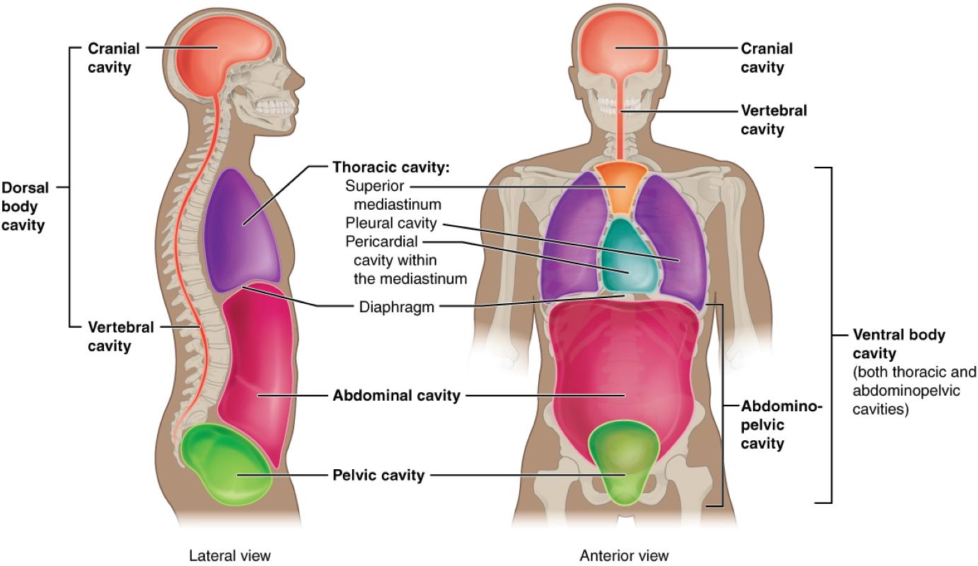
The human body is organized into distinct cavities that house and protect vital organs, with the dorsal and ventral cavities playing central roles. This image showcases the Cranial Cavity, Vertebral Cavity, Thoracic Cavity, Mediastinum, Pleural Cavity, Pericardial Cavity, Diaphragm, Abdominal Cavity, Pelvic Cavity, and Ventral Body Cavity, offering a clear view from both lateral and anterior perspectives. Exploring these cavities provides a deeper appreciation of how the body’s internal structures are safeguarded and supported.
Label Introductions:
- Cranial Cavity: The cranial cavity encases the brain, providing a protective bony structure within the skull. It cushions the brain against trauma and maintains its position for optimal function.
- Vertebral Cavity: The vertebral cavity houses the spinal cord, running through the vertebral column for protection. It allows for flexibility while shielding the spinal cord from injury.
- Dorsal Body Cavity: The dorsal body cavity encompasses the cranial and vertebral cavities, protecting the central nervous system. It is located along the posterior aspect of the body, ensuring stability.
- Thoracic Cavity: The thoracic cavity contains the heart, lungs, and major blood vessels, enclosed by the rib cage. It is separated from the abdominal cavity by the diaphragm, aiding in respiration and circulation.
- Mediastinum: The mediastinum is the central compartment of the thoracic cavity, housing the heart, trachea, and esophagus. It provides structural support and separates the lungs within the chest.
- Pleural Cavity: The pleural cavity surrounds each lung, containing a thin layer of fluid to reduce friction during breathing. It plays a critical role in maintaining lung expansion and protection.
- Pericardial Cavity: The pericardial cavity encloses the heart, providing a lubricated space to minimize friction during heartbeats. It safeguards the heart while allowing its rhythmic movements.
- Diaphragm: The diaphragm is a muscular partition separating the thoracic and abdominal cavities, essential for breathing. It contracts and relaxes to facilitate inhalation and exhalation.
- Abdominal Cavity: The abdominal cavity contains organs like the stomach, liver, and intestines, protected by the abdominal wall. It supports digestion and nutrient absorption processes.
- Pelvic Cavity: The pelvic cavity houses the bladder, reproductive organs, and lower intestines, nestled within the pelvic bones. It provides stability and protection for these essential structures.
- Ventral Body Cavity: The ventral body cavity includes the thoracic and abdominopelvic cavities, encompassing most major organs. It is located along the anterior body, facilitating organ function and accessibility.
Overview of Body Cavities
Body cavities are specialized spaces that protect and organize the body’s internal organs, ensuring their proper function. The dorsal body cavity safeguards the nervous system, while the ventral body cavity supports vital organs involved in respiration, digestion, and reproduction. Understanding these cavities is fundamental for anatomical studies and medical procedures.
- Serve as protective housings for delicate organs like the brain and heart.
- Allow for the expansion and movement of organs during physiological processes.
- Are lined with membranes that reduce friction and prevent infection.
- Enable medical professionals to locate and treat internal conditions accurately.
Dorsal Body Cavity: Protecting the Nervous System
The dorsal body cavity is a critical region that shields the central nervous system. The cranial cavity houses the brain, surrounded by the skull to prevent damage, while the vertebral cavity protects the spinal cord within the spine. This cavity’s robust structure ensures the nervous system’s integrity.
- The cranial cavity is filled with cerebrospinal fluid to cushion the brain.
- The vertebral cavity supports the spinal cord’s role in reflex actions.
- Both cavities are reinforced by bony structures for maximum protection.
- Damage to this cavity can lead to severe neurological impairments.
Ventral Body Cavity: Housing Vital Organs
The ventral body cavity is a larger space that includes the thoracic and abdominopelvic regions. The thoracic cavity contains the heart and lungs, separated by the mediastinum, while the abdominal and pelvic cavities hold digestive and reproductive organs. This cavity’s accessibility makes it key for surgical interventions.
- The pleural and pericardial cavities within the thoracic region support lung and heart function.
- The diaphragm’s movement drives the ventilation process in the thoracic cavity.
- The abdominal cavity hosts the liver, which detoxifies blood and produces bile.
- The pelvic cavity protects the bladder, which stores urine before excretion.
Thoracic Cavity: Center of Respiration and Circulation
The thoracic cavity is a dynamic space that supports the lungs and heart. The pleural cavities ensure smooth lung movement, while the pericardial cavity protects the heart’s beating action. The mediastinum organizes these structures, maintaining their spatial relationships.
- The lungs expand into the pleural cavities during inhalation.
- The heart’s position in the pericardial cavity optimizes blood flow.
- The mediastinum contains the thymus, aiding immune function.
- Injuries here can affect breathing and cardiovascular health.
Abdominopelvic Cavity: Digestive and Reproductive Hub
The abdominopelvic cavity, comprising the abdominal and pelvic cavities, is essential for digestion and reproduction. The abdominal cavity includes the stomach and intestines, while the pelvic cavity houses the bladder and reproductive organs. This region’s organs work together to sustain metabolic and excretory functions.
- The liver in the abdominal cavity produces T3 and T4 precursors indirectly via metabolism.
- The pelvic cavity supports the uterus, crucial for fetal development.
- The abdominal cavity’s peritoneum reduces friction among organs.
- Disorders here can impact nutrient absorption or urinary function.
Conclusion
The study of the dorsal body cavity and ventral body cavity reveals the intricate organization of the human body’s internal landscape. From the protective cranial and vertebral cavities to the functional thoracic, abdominal, and pelvic cavities, each plays a unique role in maintaining health. Gaining insight into these cavities enhances the ability to diagnose and treat conditions, underscoring their importance in medical practice.

