Explore the intricate process of deglutition with this detailed diagram, illustrating the voluntary and two involuntary phases: the pharyngeal phase and the esophageal phase. Learn how coordinated muscular contractions ensure the safe and efficient transport of a food bolus from the mouth to the stomach, a critical function for both nutrition and airway protection.
Swallowing, or deglutition, is a remarkably complex physiological process that seamlessly transitions from conscious initiation to an intricate series of involuntary muscular actions. It is a vital function for survival, ensuring that food and liquids are safely transported from the oral cavity to the stomach, while simultaneously protecting the respiratory airway. This overview elucidates the distinct phases of deglutition, highlighting the precise coordination of muscle contractions and structural movements that underpin this essential act.
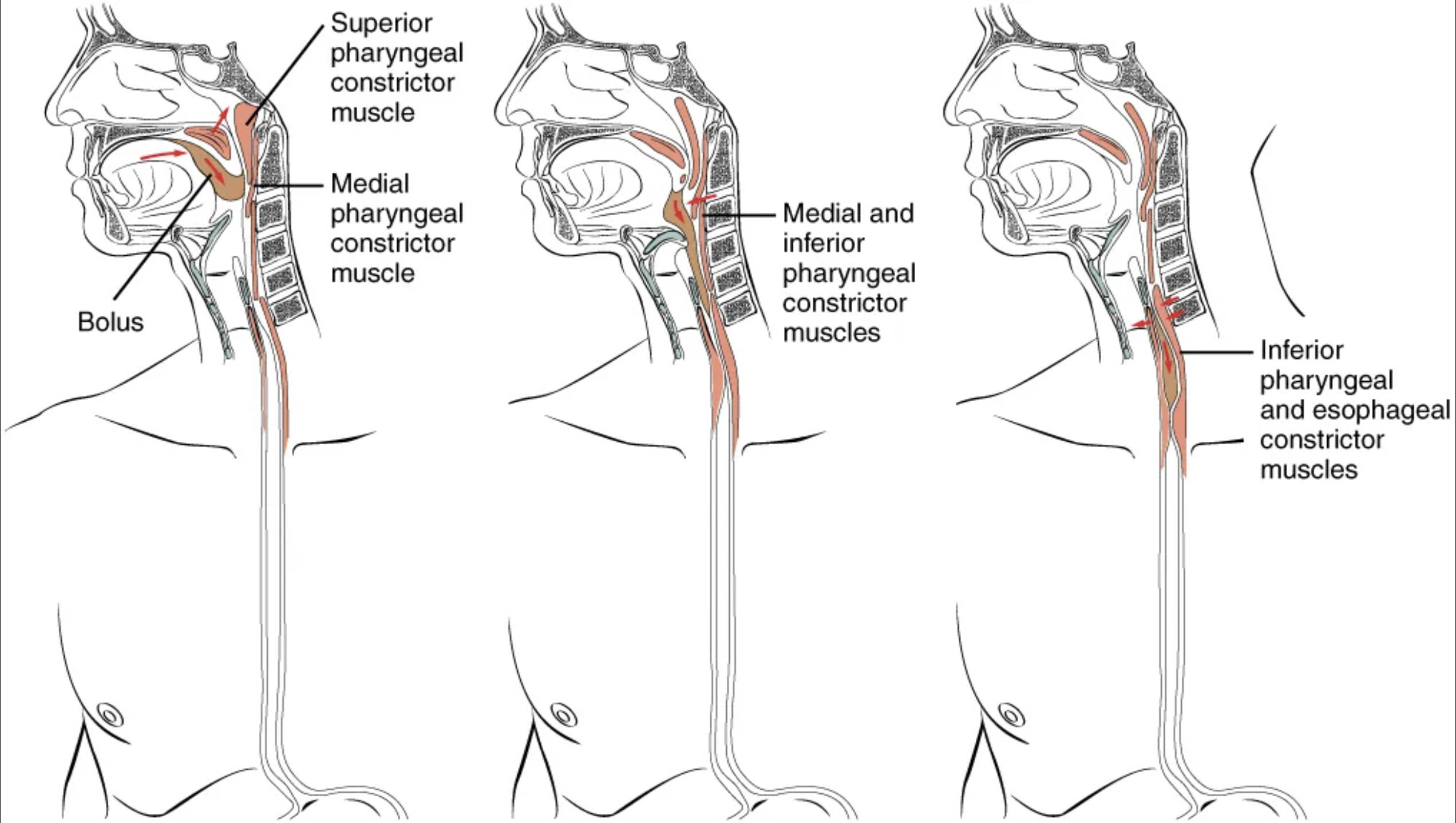
Bolus: A bolus is a soft, rounded mass of chewed food mixed with saliva, prepared in the mouth for swallowing. Its formation is the result of mechanical and chemical digestion in the oral cavity.
Superior pharyngeal constrictor muscle: This muscle is located at the uppermost part of the pharynx. During the pharyngeal phase of swallowing, it contracts to propel the food bolus downwards into the oropharynx, preventing it from moving upwards into the nasopharynx.
Medial pharyngeal constrictor muscle: Positioned below the superior pharyngeal constrictor, this muscle also contracts during the pharyngeal phase of swallowing. Its action, in concert with the other constrictors, effectively squeezes the bolus further down the pharynx towards the esophagus.
Medial and inferior pharyngeal constrictor muscles: As the bolus descends, the medial and inferior pharyngeal constrictor muscles engage in a coordinated contraction. This sequential contraction creates a wave-like action that actively pushes the bolus through the lower part of the pharynx.
Inferior pharyngeal and esophageal constrictor muscles: At the final stage of the pharyngeal phase and the beginning of the esophageal phase, these muscles continue the propulsive action. Their contraction ensures the bolus effectively transitions from the pharynx into the upper esophagus, overcoming the resistance of the upper esophageal sphincter.
The Three Phases of Swallowing
Deglutition is not a single, simple action but a highly integrated process divided into three distinct phases: the voluntary (oral) phase, the involuntary pharyngeal phase, and the involuntary esophageal phase. Each phase is critical for the safe and efficient transfer of food from the mouth to the stomach, preventing aspiration and facilitating digestion.
The three phases of swallowing are:
- Voluntary (Oral) Phase: This initial stage is conscious and involves the preparation of the food bolus. The tongue pushes the bolus against the hard palate and then posteriorly towards the oropharynx.
- Pharyngeal Phase: Once the bolus reaches the oropharynx, this involuntary phase begins, characterized by rapid and complex reflexes. Airway protection is paramount in this stage.
- Esophageal Phase: This entirely involuntary phase involves the transport of the bolus through the esophagus to the stomach via peristalsis.
During the pharyngeal phase, several critical events occur simultaneously and rapidly. As the bolus enters the oropharynx, the soft palate elevates to seal off the nasopharynx, preventing food from entering the nasal cavity. Concurrently, the larynx moves upwards and forwards, and the epiglottis folds down to cover the glottis, effectively sealing off the entrance to the trachea and protecting the airway. The pharyngeal constrictor muscles (superior, medial, and inferior) then contract in a sequential, wave-like fashion, actively propelling the food bolus downwards into the esophagus. This precise coordination ensures that food is directed away from the respiratory tract.
Following the pharyngeal phase, the esophageal phase commences as the bolus passes through the upper esophageal sphincter. This entire phase is involuntary and is driven by peristalsis, a series of rhythmic muscular contractions and relaxations that create a wave-like motion along the esophageal tube. These peristaltic waves efficiently push the bolus towards the stomach. The presence of food in the esophagus stimulates these contractions, ensuring that the bolus moves unidirectionally and reaches the lower esophageal sphincter. This sphincter then relaxes to allow the bolus into the stomach, before contracting again to prevent gastric reflux. Disruptions in any of these phases, such as dysphagia (difficulty swallowing) due to neurological conditions, muscular weakness, or structural abnormalities, can lead to serious health complications like aspiration pneumonia or malnutrition, underscoring the vital importance of this intricate physiological process.
In summary, deglutition is a finely tuned process that exemplifies the intricate coordination of the human body. From the conscious preparation of food in the oral cavity to the involuntary, sequential muscle contractions of the pharynx and esophagus, every step is critical for safe and effective nutrient intake. A comprehensive understanding of these phases is essential for medical professionals in diagnosing and treating swallowing disorders, ultimately contributing to improved patient outcomes and quality of life.

