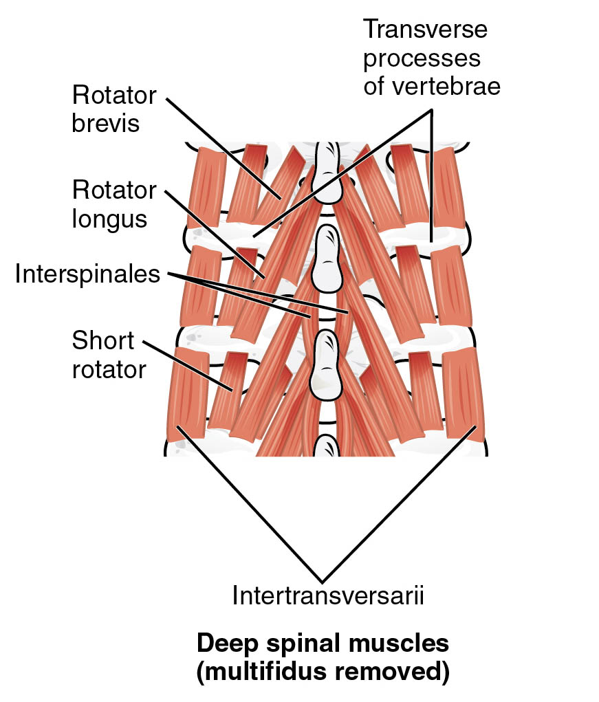
The human spine is a complex structure supported by a network of deep spinal muscles, which play a critical role in maintaining posture and facilitating movement. This anatomical image provides a detailed view of the deep spinal muscles with the multifidus removed, highlighting the intricate arrangement of muscles that stabilize the vertebral column. Exploring this illustration offers valuable insights into the musculoskeletal system, aiding in the study of spinal health and potential therapeutic approaches.
Label Introduction
- Transverse processes of vertebrae
These are bony projections extending laterally from the vertebrae, serving as attachment points for muscles and ligaments. They play a crucial role in providing structural support and enabling the range of motion in the spine. - Rotator brevis
This short muscle connects adjacent vertebrae, assisting in the rotation and stabilization of the spinal column. Its precise positioning allows for fine-tuned control during movements like twisting. - Rotator longus
A longer muscle that spans multiple vertebral levels, it contributes to rotational movements and helps maintain spinal alignment. Its extended reach enhances the overall stability of the vertebral structure. - Interspinales
These small muscles run between the spinous processes of adjacent vertebrae, aiding in the extension and stabilization of the spine. They are essential for maintaining proper posture during various activities. - Short rotator
This muscle group works in conjunction with other rotators to facilitate small, controlled rotational movements of the spine. Its action is vital for the coordination of complex spinal motions. - Intertransversarii
Located between the transverse processes of vertebrae, these muscles assist in lateral flexion and stabilize the spine during side-to-side movements. They contribute to the overall flexibility and strength of the vertebral column.
Detailed SEO Article
Overview of Deep Spinal Muscles Anatomy
Delving into the anatomy of the deep spinal muscles reveals their importance in supporting the spine’s intricate framework. This image, with the multifidus removed, showcases the underlying muscle layers that are critical for spinal stability and movement. Understanding these structures is fundamental for grasping how the body maintains posture and responds to physical demands.
- Provides a clear view of muscles like rotator brevis and rotator longus, which are pivotal for rotational stability.
- Highlights the role of interspinales and intertransversarii in fine-tuning spinal movements.
- Offers insight into the transverse processes of vertebrae as key anchoring points for these muscles.
- Demonstrates the complexity of the short rotator in coordinating subtle spinal adjustments.
Functions of Deep Spinal Muscles
The deep spinal muscles are integral to the body’s ability to perform a wide range of motions while protecting the spinal cord. Each muscle group has a specialized function that contributes to the overall health of the musculoskeletal system.
- Rotator brevis enhances rotational control, ensuring smooth and controlled turns of the torso.
- Rotator longus supports extended rotational movements, bridging multiple vertebral segments for stability.
- Interspinales assist in extending the spine, particularly during upright posture or backward bending.
- Short rotator facilitates precise adjustments, crucial for activities requiring fine motor skills.
- Intertransversarii enable lateral flexion, allowing the spine to bend sideways effectively.
- Transverse processes of vertebrae serve as robust attachment sites, distributing mechanical stress across the spine.
Clinical Relevance of Deep Spinal Muscles
The deep spinal muscles are often implicated in conditions affecting spinal health, making their study essential for therapeutic interventions. Proper functioning of these muscles can prevent or alleviate issues related to poor posture or injury.
- Weakness in rotator brevis or rotator longus may lead to reduced rotational capacity, impacting daily activities.
- Strain on interspinales can contribute to lower back pain, necessitating targeted strengthening exercises.
- Dysfunction in intertransversarii might affect lateral stability, requiring physical therapy to restore balance.
- The transverse processes of vertebrae can become stress points in cases of trauma, influencing muscle attachment integrity.
- Regular assessment of short rotator function aids in diagnosing subtle spinal misalignments.
Physical Examination and Therapeutic Approaches
Evaluating the deep spinal muscles involves a systematic approach to assess their strength and flexibility. Therapeutic techniques can target these muscles to enhance spinal health and prevent chronic issues.
- Palpation of the transverse processes of vertebrae helps identify areas of tenderness or misalignment.
- Strengthening exercises for rotator brevis and rotator longus improve rotational strength and endurance.
- Stretching routines targeting interspinales can relieve tension and improve spinal extension.
- Physical therapy for intertransversarii focuses on enhancing lateral flexibility and reducing stiffness.
- Rehabilitation programs often include short rotator exercises to support overall spinal coordination.
Anatomical Considerations for Spinal Health
A thorough understanding of the deep spinal muscles’ anatomy is crucial for maintaining long-term spinal health. The removal of multifidus in this image allows for a clearer view of these underlying structures, offering a unique perspective on their roles.
- The transverse processes of vertebrae act as critical landmarks for surgical or diagnostic procedures.
- Balanced development of rotator brevis and rotator longus supports ergonomic posture during prolonged sitting.
- Healthy interspinales contribute to resilience against degenerative changes in the spine.
- Optimal function of intertransversarii is vital for athletes engaging in lateral movements.
- The short rotator plays a subtle yet significant role in preventing compensatory movements that lead to strain.
Conclusion
The deep spinal muscles, as illustrated with the multifidus removed, form a sophisticated network that underpins spinal stability and mobility. This anatomical image serves as an invaluable resource for studying the interplay between muscles like rotator brevis, rotator longus, interspinales, short rotator, and intertransversarii, all anchored by the transverse processes of vertebrae. By exploring their functions and clinical relevance, individuals can better appreciate the importance of maintaining spinal health through targeted exercises and professional guidance. This knowledge empowers a proactive approach to preventing spinal issues and enhancing overall physical well-being.

